4J93 | pdb_00004j93

Crystal Structure of the N-Terminal Domain of HIV-1 Capsid in Complex With Inhibitor BI-1


Experimental Data Snapshot

  • Method: X-RAY DIFFRACTION
  • Resolution: 1.74 Å
  • R-Value Free: 
    0.233 (Depositor), 0.228 (DCC) 
  • R-Value Work: 
    0.193 (Depositor), 0.190 (DCC) 
  • R-Value Observed: 
    0.197 (Depositor) 

wwPDB Validation 3D Report Full Report

Validation slider image for 4J93

Ligand Structure Quality Assessment 


This is version 1.3 of the entry. See complete history

Literature

Discovery of Novel Small-Molecule HIV-1 Replication Inhibitors That Stabilize Capsid Complexes.

Lamorte, L.Titolo, S.Lemke, C.T.Goudreau, N.Mercier, J.F.Wardrop, E.Shah, V.B.von Schwedler, U.K.Langelier, C.Banik, S.S.Aiken, C.Sundquist, W.I.Mason, S.W.

(2013) Antimicrob Agents Chemother 57: 4622-4631

  • DOI: https://doi.org/10.1128/AAC.00985-13
  • Primary Citation Related Structures: 
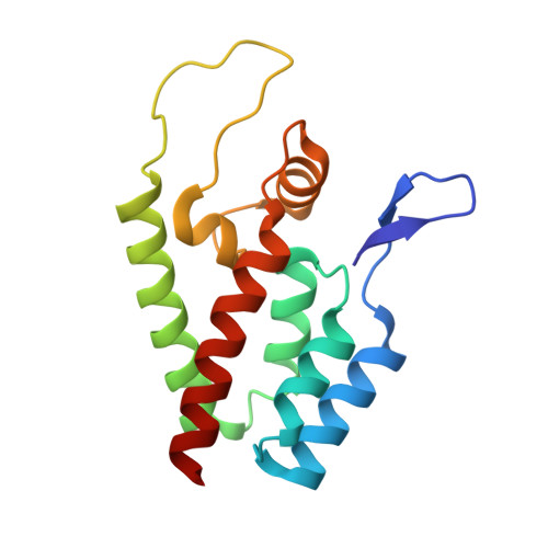
    4J93

  • PubMed Abstract: 

    The identification of novel antiretroviral agents is required to provide alternative treatment options for HIV-1-infected patients. The screening of a phenotypic cell-based viral replication assay led to the identification of a novel class of 4,5-dihydro-1H-pyrrolo[3,4-c]pyrazol-6-one (pyrrolopyrazolone) HIV-1 inhibitors, exemplified by two compounds: BI-1 and BI-2. These compounds inhibited early postentry stages of viral replication at a step(s) following reverse transcription but prior to 2 long terminal repeat (2-LTR) circle formation, suggesting that they may block nuclear targeting of the preintegration complex. Selection of viruses resistant to BI-2 revealed that substitutions at residues A105 and T107 within the capsid (CA) amino-terminal domain (CANTD) conferred high-level resistance to both compounds, implicating CA as the antiviral target. Direct binding of BI-1 and/or BI-2 to CANTD was demonstrated using isothermal titration calorimetry and nuclear magnetic resonance (NMR) chemical shift titration analyses. A high-resolution crystal structure of the BI-1:CANTD complex revealed that the inhibitor bound within a recently identified inhibitor binding pocket (CANTD site 2) between CA helices 4, 5, and 7, on the surface of the CANTD, that also corresponds to the binding site for the host factor CPSF-6. The functional consequences of BI-1 and BI-2 binding differ from previously characterized inhibitors that bind the same site since the BI compounds did not inhibit reverse transcription but stabilized preassembled CA complexes. Hence, this new class of antiviral compounds binds CA and may inhibit viral replication by stabilizing the viral capsid.


  • Organizational Affiliation
    • Department of Biological Sciences.

Macromolecule Content 

  • Total Structure Weight: 17 kDa 
  • Atom Count: 1,343 
  • Modeled Residue Count: 146 
  • Deposited Residue Count: 146 
  • Unique protein chains: 1

Macromolecules

Find similar proteins by:|  3D Structure
Entity ID: 1
MoleculeChains  Sequence LengthOrganismDetailsImage
Gag protein146Human immunodeficiency virus 1Mutation(s): 0 
Gene Names: gag
UniProt
Find proteins for Q79791 (Human immunodeficiency virus type 1 group M subtype B (isolate NY5))
Explore Q79791 
Go to UniProtKB:  Q79791
Entity Groups
Sequence Clusters30% Identity50% Identity70% Identity90% Identity95% Identity100% Identity
UniProt GroupQ79791
Sequence Annotations
Expand
Reference Sequence

Small Molecules

Ligands 2 Unique
IDChains Name / Formula / InChI Key2D Diagram3D Interactions
0OE

Query on 0OE


Download:Ideal Coordinates CCD File
B [auth A]4-{2-[5-(3-chlorophenyl)-1H-pyrazol-4-yl]-1-[3-(1H-imidazol-1-yl)propyl]-1H-benzimidazol-5-yl}benzoic acid
C29 H23 Cl N6 O2
LGXPZODGTPYYBU-UHFFFAOYSA-N
1JR

Query on 1JR


Download:Ideal Coordinates CCD File
C [auth A](4S)-3-phenyl-4-(pyridin-3-yl)-4,5-dihydropyrrolo[3,4-c]pyrazol-6(2H)-one
C16 H12 N4 O
OHFQLOFZGKOEBH-ZDUSSCGKSA-N

Experimental Data & Validation

Experimental Data

  • Method: X-RAY DIFFRACTION
  • Resolution: 1.74 Å
  • R-Value Free:  0.233 (Depositor), 0.228 (DCC) 
  • R-Value Work:  0.193 (Depositor), 0.190 (DCC) 
  • R-Value Observed: 0.197 (Depositor) 
Space Group: P 21 21 21
Unit Cell:
Length ( Å )Angle ( ˚ )
a = 40.779α = 90
b = 42.494β = 90
c = 83.279γ = 90
Software Package:
Software NamePurpose
MAR345dtbdata collection
PHENIXmodel building
PHENIXrefinement
HKL-2000data reduction
HKL-2000data scaling
PHENIXphasing

Structure Validation

View Full Validation Report


Ligand Structure Quality Assessment 


Entry History 

Deposition Data

  • Released Date: 2013-07-10 
  • Deposition Author(s): Lemke, C.T.

Revision History  (Full details and data files)

  • Version 1.0: 2013-07-10
    Type: Initial release
  • Version 1.1: 2013-07-17
    Changes: Database references
  • Version 1.2: 2013-10-09
    Changes: Database references
  • Version 1.3: 2024-02-28
    Changes: Data collection, Database references, Derived calculations