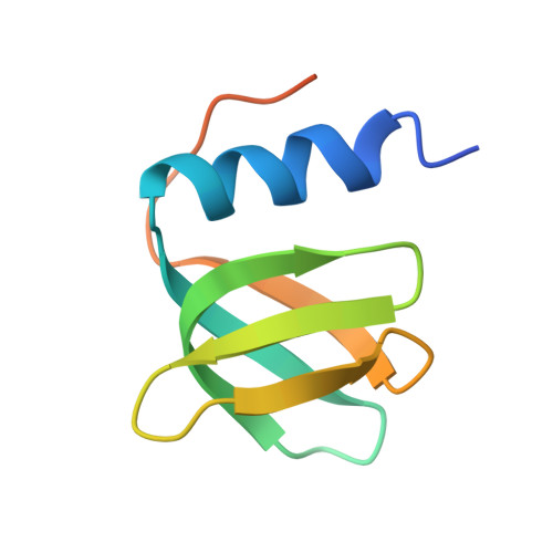
4J5Y

Crystal structure of Hfq from Pseudomonas aeruginosa in complex with ATP


Experimental Data Snapshot

  • Method: X-RAY DIFFRACTION
  • Resolution: 2.10 Å
  • R-Value Free: 0.252 
  • R-Value Work: 0.184 
  • R-Value Observed: 0.188 

Starting Model: experimental
View more details

wwPDB Validation   3D Report Full Report


Ligand Structure Quality Assessment 


This is version 1.2 of the entry. See complete history


Literature

Hfq binds ribonucleotides in three different RNA-binding sites.

Murina, V.Lekontseva, N.Nikulin, A.

(2013) Acta Crystallogr D Biol Crystallogr 69: 1504-1513

  • DOI: https://doi.org/10.1107/S090744491301010X
  • Primary Citation of Related Structures:  
    3QUI, 4J5Y, 4J6W, 4J6X, 4J6Y

  • PubMed Abstract: 

    The Hfq protein forms a doughnut-shaped homohexamer that possesses RNA-binding activity. There are two distinct RNA-binding surfaces located on the proximal and the distal sides of the hexamer. The proximal side is involved in the binding of mRNA and small noncoding RNAs (sRNAs), while the distal side has an affinity for A-rich RNA sequences. In this work, the ability of various ribonucleotides to form complexes with Hfq from Pseudomonas aeruginosa has been tested using X-ray crystallography. ATP and ADPNP have been located in the distal R-site, which is a site for poly(A) RNA binding. UTP has been found in the so-called lateral RNA-binding site at the proximal surface. CTP has been found in both the distal R-site and the proximal U-binding site. GTP did not form a complex with Hfq under the conditions tested. The results have demonstrated the power of the crystallographic method for locating ribonucleotides and predicting single-stranded RNA-binding sites on the protein surface.


  • Organizational Affiliation

    Institute of Protein Research, RAS, Institutskaya 4, Pushchino 142290, Moscow Region, Russian Federation.


Macromolecules
Find similar proteins by:  (by identity cutoff)  |  3D Structure
Entity ID: 1
MoleculeChains Sequence LengthOrganismDetailsImage
Protein hfq
A, B, C, D, E
A, B, C, D, E, F
82Pseudomonas aeruginosa PAO1Mutation(s): 0 
Gene Names: hfqPA4944
UniProt
Find proteins for Q9HUM0 (Pseudomonas aeruginosa (strain ATCC 15692 / DSM 22644 / CIP 104116 / JCM 14847 / LMG 12228 / 1C / PRS 101 / PAO1))
Explore Q9HUM0 
Go to UniProtKB:  Q9HUM0
Entity Groups  
Sequence Clusters30% Identity50% Identity70% Identity90% Identity95% Identity100% Identity
UniProt GroupQ9HUM0
Sequence Annotations
Expand
  • Reference Sequence
Small Molecules
Ligands 3 Unique
IDChains Name / Formula / InChI Key2D Diagram3D Interactions
ATP
Query on ATP

Download Ideal Coordinates CCD File 
G [auth A],
I [auth B],
J [auth C],
K [auth D],
N [auth F]
ADENOSINE-5'-TRIPHOSPHATE
C10 H16 N5 O13 P3
ZKHQWZAMYRWXGA-KQYNXXCUSA-N
PEG
Query on PEG

Download Ideal Coordinates CCD File 
L [auth D]DI(HYDROXYETHYL)ETHER
C4 H10 O3
MTHSVFCYNBDYFN-UHFFFAOYSA-N
NA
Query on NA

Download Ideal Coordinates CCD File 
H [auth A],
M [auth D]
SODIUM ION
Na
FKNQFGJONOIPTF-UHFFFAOYSA-N
Experimental Data & Validation

Experimental Data

  • Method: X-RAY DIFFRACTION
  • Resolution: 2.10 Å
  • R-Value Free: 0.252 
  • R-Value Work: 0.184 
  • R-Value Observed: 0.188 
  • Space Group: P 21 21 21
Unit Cell:
Length ( Å )Angle ( ˚ )
a = 61.45α = 90
b = 73.68β = 90
c = 107.56γ = 90
Software Package:
Software NamePurpose
PROTEUM PLUSdata collection
PHENIXmodel building
PHENIXrefinement
PROTEUM PLUSdata reduction
PROTEUM PLUSdata scaling
PHENIXphasing

Structure Validation

View Full Validation Report


Ligand Structure Quality Assessment 


Entry History 

Deposition Data

Revision History  (Full details and data files)

  • Version 1.0: 2013-07-31
    Type: Initial release
  • Version 1.1: 2013-08-21
    Changes: Database references
  • Version 1.2: 2023-09-20
    Changes: Data collection, Database references, Derived calculations, Refinement description