4J47 | pdb_00004j47

Crystal structure of XIAP-BIR2 domain with SVPI bound


Experimental Data Snapshot

  • Method: X-RAY DIFFRACTION
  • Resolution: 1.35 Å
  • R-Value Free: 
    0.194 (Depositor), 0.190 (DCC) 
  • R-Value Work: 
    0.174 (Depositor), 0.183 (DCC) 
  • R-Value Observed: 
    0.175 (Depositor) 

Starting Model: experimental
View more details

wwPDB Validation 3D Report Full Report

Validation slider image for 4J47

This is version 1.2 of the entry. See complete history

Literature

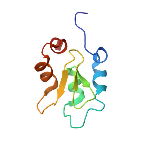
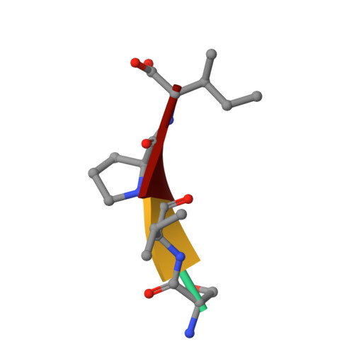
The structure of XIAP BIR2: understanding the selectivity of the BIR domains.

Lukacs, C.Belunis, C.Crowther, R.Danho, W.Gao, L.Goggin, B.Janson, C.A.Li, S.Remiszewski, S.Schutt, A.Thakur, M.K.Singh, S.K.Swaminathan, S.Pandey, R.Tyagi, R.Gosu, R.Kamath, A.V.Kuglstatter, A.

(2013) Acta Crystallogr D Biol Crystallogr 69: 1717-1725

  • DOI: https://doi.org/10.1107/S0907444913016284
  • Primary Citation Related Structures: 
    4J3Y, 4J44, 4J45, 4J46, 4J47, 4J48

  • PubMed Abstract: 

    XIAP, a member of the inhibitor of apoptosis family of proteins, is a critical regulator of apoptosis. Inhibition of the BIR domain-caspase interaction is a promising approach towards treating cancer. Previous work has been directed towards inhibiting the BIR3-caspase-9 interaction, which blocks the intrinsic apoptotic pathway; selectively inhibiting the BIR2-caspase-3 interaction would also block the extrinsic pathway. The BIR2 domain of XIAP has successfully been crystallized; peptides and small-molecule inhibitors can be soaked into these crystals, which diffract to high resolution. Here, the BIR2 apo crystal structure and the structures of five BIR2-tetrapeptide complexes are described. The structural flexibility observed on comparing these structures, along with a comparison with XIAP BIR3, affords an understanding of the structural elements that drive selectivity between BIR2 and BIR3 and which can be used to design BIR2-selective inhibitors.


  • Organizational Affiliation
    • Discovery Technologies, Hoffmann-La Roche, 340 Kingsland Street, Nutley, NJ 07110, USA.

Macromolecule Content 

  • Total Structure Weight: 20.42 kDa 
  • Atom Count: 1,487 
  • Modeled Residue Count: 163 
  • Deposited Residue Count: 176 
  • Unique protein chains: 2

Macromolecules

Find similar proteins by:|  3D Structure
Entity ID: 1
MoleculeChains  Sequence LengthOrganismDetailsImage
E3 ubiquitin-protein ligase XIAP
A, C
86Homo sapiensMutation(s): 2 
Gene Names: API3BIRC4IAP3U32974XIAP
EC: 6.3.2 (PDB Primary Data), 2.3.2.27 (UniProt)
UniProt & NIH Common Fund Data Resources
Find proteins for P98170 (Homo sapiens)
Explore P98170 
Go to UniProtKB:  P98170
PHAROS:  P98170
GTEx:  ENSG00000101966 
Entity Groups
Sequence Clusters30% Identity50% Identity70% Identity90% Identity95% Identity100% Identity
UniProt GroupP98170
Sequence Annotations
Expand
Reference Sequence
Find similar proteins by:  Sequence   |   3D Structure  
Entity ID: 2
MoleculeChains  Sequence LengthOrganismDetailsImage
PEPTIDE (SER-VAL-PRO-ILE)4Homo sapiensMutation(s): 0 

Experimental Data & Validation

Experimental Data

  • Method: X-RAY DIFFRACTION
  • Resolution: 1.35 Å
  • R-Value Free:  0.194 (Depositor), 0.190 (DCC) 
  • R-Value Work:  0.174 (Depositor), 0.183 (DCC) 
  • R-Value Observed: 0.175 (Depositor) 
Space Group: I 4 2 2
Unit Cell:
Length ( Å )Angle ( ˚ )
a = 74.539α = 90
b = 74.539β = 90
c = 108.671γ = 90
Software Package:
Software NamePurpose
REFMACrefinement
XDSdata reduction
SCALAdata scaling
CNXrefinement

Structure Validation

View Full Validation Report


Entry History 

Deposition Data

Revision History  (Full details and data files)

  • Version 1.0: 2013-09-25
    Type: Initial release
  • Version 1.1: 2019-07-17
    Changes: Data collection, Refinement description
  • Version 1.2: 2023-09-20
    Changes: Data collection, Database references, Derived calculations, Refinement description