Crystal structure of a UDP-GlcNAc epimerase for surface polysaccharide biosynthesis in Acinetobacter baumannii.
Shah, B.S., Ashwood, H.E., Harrop, S.J., Farrugia, D.N., Paulsen, I.T., Mabbutt, B.C.(2018) PLoS One 13: e0191610-e0191610
- PubMed: 29352301 Search on PubMedSearch on PubMed Central
- DOI: https://doi.org/10.1371/journal.pone.0191610
- Primary Citation Related Structures:
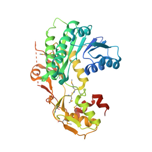
4J2O - PubMed Abstract:
With new strains of Acinetobacter baumannii undergoing genomic analysis, it has been possible to define regions of genomic plasticity (RGPs), encoding specific adaptive elements. For a selected RGP from a community-derived isolate of A. baumannii, we outline sequences compatible with biosynthetic machinery of surface polysaccharides, specifically enzymes utilized in the dehydration and conversion of UDP-N-acetyl-D-glucosamine (UDP-D-GlcNAc). We have determined the crystal structure of one of these, the epimerase Ab-WbjB. This dehydratase belongs to the 'extended' short-chain dehydrogenase/reductase (SDR) family, related in fold to previously characterised enzymes CapE and FlaA1. Our 2.65Å resolution structure of Ab-WbjB shows a hexamer, organised into a trimer of chain pairs, with coenzyme NADP+ occupying each chain. Specific active-site interactions between each coenzyme and a lysine quaternary group of a neighbouring chain interconnect adjacent dimers, so stabilising the hexameric form. We show UDP-GlcNAc to be a specific substrate for Ab-WbjB, with binding evident by ITC (Ka = 0.23 μmol-1). The sequence of Ab-WbjB shows variation from the consensus active-site motifs of many SDR enzymes, demonstrating a likely catalytic role for a specific threonine sidechain (as an alternative to tyrosine) in the canonical active site chemistry of these epimerases.
- Department of Molecular Sciences, Macquarie University, Sydney, Australia.
Organizational Affiliation: