4J12 | pdb_00004j12

monomeric Fc


Experimental Data Snapshot

  • Method: X-RAY DIFFRACTION
  • Resolution: 1.90 Å
  • R-Value Free: 
    0.238 (Depositor), 0.249 (DCC) 
  • R-Value Work: 
    0.222 (Depositor), 0.234 (DCC) 
  • R-Value Observed: 
    0.223 (Depositor) 

wwPDB Validation 3D Report Full Report

Validation slider image for 4J12

Ligand Structure Quality Assessment 


This is version 2.1 of the entry. See complete history

Literature

Engineering a Monomeric Fc Domain Modality by N-Glycosylation for the Half-life Extension of Biotherapeutics.

Ishino, T.Wang, M.Mosyak, L.Tam, A.Duan, W.Svenson, K.Joyce, A.O'Hara, D.M.Lin, L.Somers, W.S.Kriz, R.

(2013) J Biological Chem 288: 16529-16537

  • DOI: https://doi.org/10.1074/jbc.M113.457689
  • Primary Citation Related Structures: 
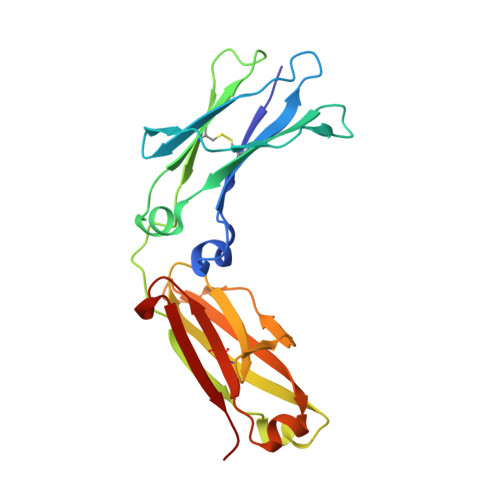
    4J12

  • PubMed Abstract: 

    Human IgG is a bivalent molecule that has two identical Fab domains connected by a dimeric Fc domain. For therapeutic purposes, however, the bivalency of IgG and Fc fusion proteins could cause undesired properties. We therefore engineered the conversion of the natural dimeric Fc domain to a highly soluble monomer by introducing two Asn-linked glycans onto the hydrophobic C(H)3-C(H)3 dimer interface. The monomeric Fc (monoFc) maintained the binding affinity for neonatal Fc receptor (FcRn) in a pH-dependent manner. We solved the crystal structure of monoFc, which explains how the carbohydrates can stabilize the protein surface and provides the rationale for molecular recognition between monoFc and FcRn. The monoFc prolonged the in vivo half-life of an antibody Fab domain, and a tandem repeat of the monoFc further prolonged the half-life. This monoFc modality can be used to improve the pharmacokinetics of monomeric therapeutic proteins with an option to modulate the degree of half-life extension.


  • Organizational Affiliation
    • From Global Biotherapeutics Technologies, Pfizer Inc., Cambridge, Massachusetts 02140. Electronic address: tetsuya.ishino@pfizer.com.

Macromolecule Content 

  • Total Structure Weight: 25.33 kDa 
  • Atom Count: 1,927 
  • Modeled Residue Count: 209 
  • Deposited Residue Count: 210 
  • Unique protein chains: 1

Macromolecules

Find similar proteins by:|  3D Structure
Entity ID: 1
MoleculeChains  Sequence LengthOrganismDetailsImage
human Fc fragment210Homo sapiensMutation(s): 3 
UniProt & NIH Common Fund Data Resources
Find proteins for P01857 (Homo sapiens)
Explore P01857 
Go to UniProtKB:  P01857
PHAROS:  P01857
GTEx:  ENSG00000211896 
Entity Groups
Sequence Clusters30% Identity50% Identity70% Identity90% Identity95% Identity100% Identity
UniProt GroupP01857
Glycosylation
Glycosylation Sites: 2Go to GlyGen: P01857-2
Sequence Annotations
Expand
Reference Sequence

Oligosaccharides

Help  
Entity ID: 2
MoleculeChains Length2D Diagram GlycosylationD Interactions
2-acetamido-2-deoxy-beta-D-glucopyranose-(1-2)-alpha-D-mannopyranose-(1-3)-[2-acetamido-2-deoxy-beta-D-glucopyranose-(1-2)-alpha-D-mannopyranose-(1-6)]beta-D-mannopyranose-(1-4)-2-acetamido-2-deoxy-beta-D-glucopyranose-(1-4)-2-acetamido-2-deoxy-beta-D-glucopyranose
B
7N-Glycosylation
Glycosylation Resources
GlyTouCan: G39213VZ
GlyCosmos: G39213VZ
GlyGen: G39213VZ

Small Molecules

Ligands 1 Unique
IDChains Name / Formula / InChI Key2D Diagram3D Interactions
NAG

Query on NAG


Download:Ideal Coordinates CCD File
C [auth A]2-acetamido-2-deoxy-beta-D-glucopyranose
C8 H15 N O6
OVRNDRQMDRJTHS-FMDGEEDCSA-N

Experimental Data & Validation

Experimental Data

  • Method: X-RAY DIFFRACTION
  • Resolution: 1.90 Å
  • R-Value Free:  0.238 (Depositor), 0.249 (DCC) 
  • R-Value Work:  0.222 (Depositor), 0.234 (DCC) 
  • R-Value Observed: 0.223 (Depositor) 
Space Group: P 31 1 2
Unit Cell:
Length ( Å )Angle ( ˚ )
a = 64.224α = 90
b = 64.224β = 90
c = 146.937γ = 120
Software Package:
Software NamePurpose
HKL-2000data collection
PHASESphasing
BUSTERrefinement
HKL-2000data reduction
HKL-2000data scaling

Structure Validation

View Full Validation Report


Ligand Structure Quality Assessment 


Entry History 

Deposition Data

Revision History  (Full details and data files)

  • Version 1.0: 2013-05-01
    Type: Initial release
  • Version 1.1: 2013-07-24
    Changes: Database references
  • Version 1.2: 2018-01-24
    Changes: Structure summary
  • Version 1.3: 2018-06-13
    Changes: Data collection, Database references
  • Version 2.0: 2020-07-29
    Type: Remediation
    Reason: Carbohydrate remediation
    Changes: Atomic model, Data collection, Database references, Derived calculations, Structure summary
  • Version 2.1: 2024-11-27
    Changes: Data collection, Database references, Structure summary