4IZG | pdb_00004izg

Crystal structure of an enolase (mandelate racemase subgroup) from paracococus denitrificans pd1222 (target nysgrc-012907) with bound cis-4oh-d-proline betaine (product)


Experimental Data Snapshot

  • Method: X-RAY DIFFRACTION
  • Resolution: 1.70 Å
  • R-Value Free: 
    0.197 (Depositor), 0.196 (DCC) 
  • R-Value Work: 
    0.165 (Depositor), 0.166 (DCC) 
  • R-Value Observed: 
    0.167 (Depositor) 

wwPDB Validation 3D Report Full Report

Validation slider image for 4IZG

Ligand Structure Quality Assessment 


This is version 1.2 of the entry. See complete history

Literature

Prediction and biochemical demonstration of a catabolic pathway for the osmoprotectant proline betaine.

Kumar, R.Zhao, S.Vetting, M.W.Wood, B.M.Sakai, A.Cho, K.Solbiati, J.Almo, S.C.Sweedler, J.V.Jacobson, M.P.Gerlt, J.A.Cronan, J.E.

(2014) mBio 5: e00933-e00913

  • DOI: https://doi.org/10.1128/mBio.00933-13
  • Primary Citation Related Structures: 
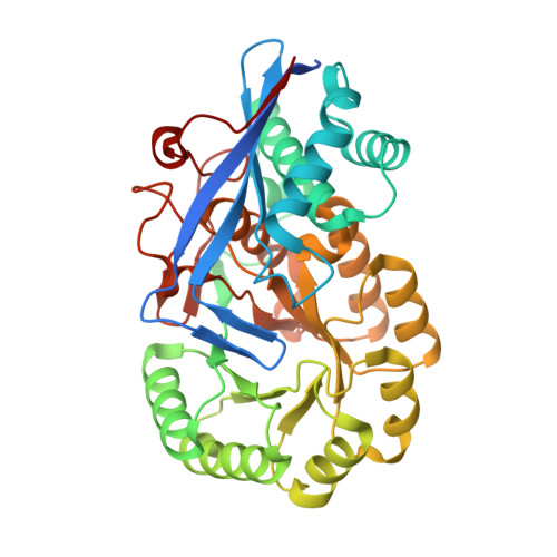
    4E8G, 4IZG, 4J1O

  • PubMed Abstract: 

    Through the use of genetic, enzymatic, metabolomic, and structural analyses, we have discovered the catabolic pathway for proline betaine, an osmoprotectant, in Paracoccus denitrificans and Rhodobacter sphaeroides. Genetic and enzymatic analyses showed that several of the key enzymes of the hydroxyproline betaine degradation pathway also function in proline betaine degradation. Metabolomic analyses detected each of the metabolic intermediates of the pathway. The proline betaine catabolic pathway was repressed by osmotic stress and cold stress, and a regulatory transcription factor was identified. We also report crystal structure complexes of the P. denitrificans HpbD hydroxyproline betaine epimerase/proline betaine racemase with l-proline betaine and cis-hydroxyproline betaine. At least half of the extant protein annotations are incorrect, and the errors propagate as the number of genome sequences increases exponentially. A large-scale, multidisciplinary sequence- and structure-based strategy for functional assignment of bacterial enzymes of unknown function has demonstrated the pathway for catabolism of the osmoprotectant proline betaine.

Macromolecule Content 

  • Total Structure Weight: 87.84 kDa 
  • Atom Count: 6,409 
  • Modeled Residue Count: 738 
  • Deposited Residue Count: 782 
  • Unique protein chains: 1

Macromolecules

Find similar proteins by:|  3D Structure
Entity ID: 1
MoleculeChains  Sequence LengthOrganismDetailsImage
Mandelate racemase/muconate lactonizing enzyme, N-terminal domain protein
A, B
391Paracoccus denitrificans PD1222Mutation(s): 0 
EC: 5.1.1.22
UniProt
Find proteins for A1B198 (Paracoccus denitrificans (strain Pd 1222))
Explore A1B198 
Go to UniProtKB:  A1B198
Entity Groups
Sequence Clusters30% Identity50% Identity70% Identity90% Identity95% Identity100% Identity
UniProt GroupA1B198
Sequence Annotations
Expand
Reference Sequence

Small Molecules

Ligands 3 Unique
IDChains Name / Formula / InChI Key2D Diagram3D Interactions
4OP

Query on 4OP


Download:Ideal Coordinates CCD File
JA [auth B],
R [auth A]
(2S,4S)-2-carboxy-4-hydroxy-1,1-dimethylpyrrolidinium
C7 H14 N O3
MUNWAHDYFVYIKH-PHDIDXHHSA-O
IOD

Query on IOD


Download:Ideal Coordinates CCD File
AA [auth B]
BA [auth B]
C [auth A]
CA [auth B]
D [auth A]
AA [auth B],
BA [auth B],
C [auth A],
CA [auth B],
D [auth A],
DA [auth B],
E [auth A],
EA [auth B],
F [auth A],
FA [auth B],
G [auth A],
GA [auth B],
H [auth A],
I [auth A],
J [auth A],
K [auth A],
L [auth A],
M [auth A],
N [auth A],
S [auth B],
T [auth B],
U [auth B],
V [auth B],
W [auth B],
X [auth B],
Y [auth B],
Z [auth B]
IODIDE ION
I
XMBWDFGMSWQBCA-UHFFFAOYSA-M
MG

Query on MG


Download:Ideal Coordinates CCD File
HA [auth B],
IA [auth B],
O [auth A],
P [auth A],
Q [auth A]
MAGNESIUM ION
Mg
JLVVSXFLKOJNIY-UHFFFAOYSA-N

Experimental Data & Validation

Experimental Data

  • Method: X-RAY DIFFRACTION
  • Resolution: 1.70 Å
  • R-Value Free:  0.197 (Depositor), 0.196 (DCC) 
  • R-Value Work:  0.165 (Depositor), 0.166 (DCC) 
  • R-Value Observed: 0.167 (Depositor) 
Space Group: P 4 21 2
Unit Cell:
Length ( Å )Angle ( ˚ )
a = 117.463α = 90
b = 117.463β = 90
c = 111.049γ = 90
Software Package:
Software NamePurpose
SCALAdata scaling
PHENIXrefinement
PDB_EXTRACTdata extraction
MOSFLMdata reduction
PHENIXphasing

Structure Validation

View Full Validation Report


Ligand Structure Quality Assessment 


Entry History 

Revision History  (Full details and data files)

  • Version 1.0: 2013-02-13
    Type: Initial release
  • Version 1.1: 2018-01-24
    Changes: Database references
  • Version 1.2: 2024-02-28
    Changes: Data collection, Database references, Derived calculations