4IVS | pdb_00004ivs

Crystal structure of BACE1 with its inhibitor


Experimental Data Snapshot

  • Method: X-RAY DIFFRACTION
  • Resolution: 2.64 Å
  • R-Value Free: 
    0.227 (Depositor), 0.185 (DCC) 
  • R-Value Work: 
    0.171 (Depositor), 0.174 (DCC) 
  • R-Value Observed: 
    0.173 (Depositor) 

Starting Model: experimental
View more details

wwPDB Validation 3D Report Full Report

Validation slider image for 4IVS

Ligand Structure Quality Assessment 


This is version 1.2 of the entry. See complete history

Literature

Virtual screening and structure-based discovery of indole acylguanidines as potent beta-secretase (BACE1) inhibitors

Zou, Y.Li, L.Chen, W.Y.Chen, T.T.Ma, L.Wang, X.Xiong, B.Xu, Y.C.Shen, J.

(2013) Molecules 18: 5706-5722

  • DOI: https://doi.org/10.3390/molecules18055706
  • Primary Citation Related Structures: 
    4IVS, 4IVT

  • PubMed Abstract: 

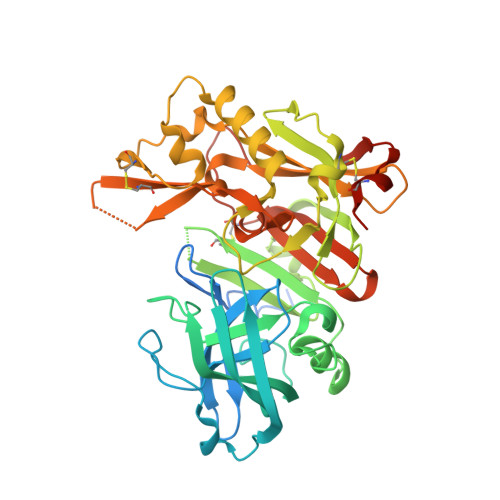
    Proteolytic cleavage of amyloid precursor protein by β-secretase (BACE1) is a key step in generating the N-terminal of β-amyloid (Aβ), which further forms into amyloid plaques that are considered as the hallmark of Alzheimer's disease. Inhibitors of BACE1 can reduce the levels of Aβ and thus have a therapeutic potential for treating the disease. We report here the identification of a series of small molecules bearing an indole acylguanidine core structure as potent BACE1 inhibitors. The initial weak fragment was discovered by virtual screening, and followed with a hit-to-lead optimization. With the aid of co-crystal structures of two discovered inhibitors (compounds 19 and 25) with BACE1, we explored the SAR around the indole and aryl groups, and obtained several BACE1 inhibitors about 1,000-fold more potent than the initial fragment hit. Accompanying the lead optimization, a previously under-explored sub-site opposite the flap loop was redefined as a potential binding site for later BACE1 inhibitor design.


  • Organizational Affiliation
    • State Key Laboratory of Drug Research, Shanghai Institute of Materia Medica, Chinese Academy of Sciences, 555 Zuchongzhi Road, Zhangjiang Hi-Tech Park, Shanghai 201203, China.

Macromolecule Content 

  • Total Structure Weight: 48.68 kDa 
  • Atom Count: 3,022 
  • Modeled Residue Count: 373 
  • Deposited Residue Count: 433 
  • Unique protein chains: 1

Macromolecules

Find similar proteins by:|  3D Structure
Entity ID: 1
MoleculeChains  Sequence LengthOrganismDetailsImage
Beta-secretase 1433Homo sapiensMutation(s): 2 
Gene Names: BACE1
EC: 3.4.23.46
UniProt & NIH Common Fund Data Resources
Find proteins for P56817 (Homo sapiens)
Explore P56817 
Go to UniProtKB:  P56817
PHAROS:  P56817
GTEx:  ENSG00000186318 
Entity Groups
Sequence Clusters30% Identity50% Identity70% Identity90% Identity95% Identity100% Identity
UniProt GroupP56817
Sequence Annotations
Expand
Reference Sequence

Small Molecules

Ligands 1 Unique
IDChains Name / Formula / InChI Key2D Diagram3D Interactions
VSI

Query on VSI


Download:Ideal Coordinates CCD File
B [auth A]N-{N-[4-(acetylamino)-3,5-dichlorobenzyl]carbamimidoyl}-2-(6-cyano-1H-indol-1-yl)acetamide
C21 H18 Cl2 N6 O2
QNSBKNRQYCQXMO-UHFFFAOYSA-N

Experimental Data & Validation

Experimental Data

  • Method: X-RAY DIFFRACTION
  • Resolution: 2.64 Å
  • R-Value Free:  0.227 (Depositor), 0.185 (DCC) 
  • R-Value Work:  0.171 (Depositor), 0.174 (DCC) 
  • R-Value Observed: 0.173 (Depositor) 
Space Group: C 2 2 21
Unit Cell:
Length ( Å )Angle ( ˚ )
a = 104.883α = 90
b = 129.376β = 90
c = 77.249γ = 90
Software Package:
Software NamePurpose
XSCALEdata scaling
PHASERphasing
PHENIXrefinement
PDB_EXTRACTdata extraction
Blu-Icedata collection

Structure Validation

View Full Validation Report


Ligand Structure Quality Assessment 


Entry History 

Deposition Data

Revision History  (Full details and data files)

  • Version 1.0: 2013-11-13
    Type: Initial release
  • Version 1.1: 2023-11-08
    Changes: Data collection, Database references, Derived calculations, Refinement description
  • Version 1.2: 2024-10-16
    Changes: Structure summary