4IOP

Crystal structure of NKp65 bound to its ligand KACL


Experimental Data Snapshot

  • Method: X-RAY DIFFRACTION
  • Resolution: 3.20 Å
  • R-Value Free: 0.297 
  • R-Value Work: 0.252 
  • R-Value Observed: 0.252 

wwPDB Validation   3D Report Full Report


This is version 2.0 of the entry. See complete history


Literature

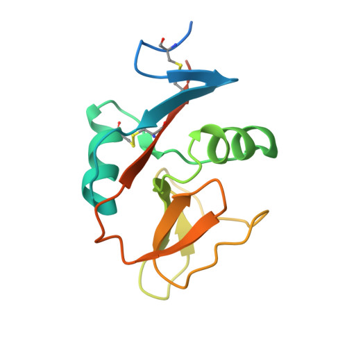
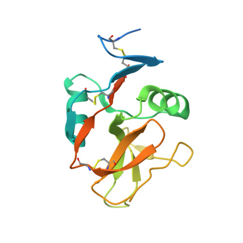
Structure of NKp65 bound to its keratinocyte ligand reveals basis for genetically linked recognition in natural killer gene complex.

Li, Y.Wang, Q.Chen, S.Brown, P.H.Mariuzza, R.A.

(2013) Proc Natl Acad Sci U S A 110: 11505-11510

  • DOI: https://doi.org/10.1073/pnas.1303300110
  • Primary Citation of Related Structures:  
    4IOP

  • PubMed Abstract: 

    The natural killer (NK) gene complex (NKC) encodes numerous C-type lectin-like receptors that govern the activity of NK cells. Although some of these receptors (Ly49s, NKG2D, CD94/NKG2A) recognize MHC or MHC-like molecules, others (Nkrp1, NKRP1A, NKp80, NKp65) instead bind C-type lectin-like ligands to which they are genetically linked in the NKC. To understand the basis for this recognition, we determined the structure of human NKp65, an activating receptor implicated in the immunosurveillance of skin, bound to its NKC-encoded ligand keratinocyte-associated C-type lectin (KACL). Whereas KACL forms a homodimer resembling other C-type lectin-like dimers, NKp65 is monomeric. The binding mode in the NKp65-KACL complex, in which a monomeric receptor engages a dimeric ligand, is completely distinct from those used by Ly49s, NKG2D, or CD94/NKG2A. The structure explains the exceptionally high affinity of the NKp65-KACL interaction compared with other cell-cell interaction pairs (KD = 6.7 × 10(-10) M), which may compensate for the monomeric nature of NKp65 to achieve cell activation. This previously unreported structure of an NKC-encoded receptor-ligand complex, coupled with mutational analysis of the interface, establishes a docking template that is directly applicable to other genetically linked pairs in the NKC, including Nkrp1-Clr, NKRP1A-LLT1, and NKp80-AICL.


  • Organizational Affiliation

    W. M. Keck Laboratory for Structural Biology, University of Maryland Institute for Bioscience and Biotechnology Research, Rockville, MD 20850, USA.


Macromolecules
Find similar proteins by:  (by identity cutoff)  |  3D Structure
Entity ID: 1
MoleculeChains Sequence LengthOrganismDetailsImage
C-type lectin domain family 2 member A142Homo sapiensMutation(s): 0 
Gene Names: CLEC2AKACLUNQ5792/PRO19597
UniProt & NIH Common Fund Data Resources
Find proteins for Q6UVW9 (Homo sapiens)
Explore Q6UVW9 
Go to UniProtKB:  Q6UVW9
PHAROS:  Q6UVW9
GTEx:  ENSG00000188393 
Entity Groups  
Sequence Clusters30% Identity50% Identity70% Identity90% Identity95% Identity100% Identity
UniProt GroupQ6UVW9
Sequence Annotations
Expand
  • Reference Sequence
Find similar proteins by:  (by identity cutoff)  |  3D Structure
Entity ID: 2
MoleculeChains Sequence LengthOrganismDetailsImage
Killer cell lectin-like receptor subfamily F member 2158Homo sapiensMutation(s): 0 
Gene Names: KLRF2
UniProt & NIH Common Fund Data Resources
Find proteins for D3W0D1 (Homo sapiens)
Explore D3W0D1 
Go to UniProtKB:  D3W0D1
PHAROS:  D3W0D1
GTEx:  ENSG00000256797 
Entity Groups  
Sequence Clusters30% Identity50% Identity70% Identity90% Identity95% Identity100% Identity
UniProt GroupD3W0D1
Sequence Annotations
Expand
  • Reference Sequence
Oligosaccharides

Help

Entity ID: 3
MoleculeChains Length2D Diagram Glycosylation3D Interactions
alpha-D-mannopyranose-(1-4)-2-acetamido-2-deoxy-beta-D-glucopyranose-(1-4)-2-acetamido-2-deoxy-beta-D-glucopyranose
C, D
3N-Glycosylation
Glycosylation Resources
GlyTouCan:  G62182OO
GlyCosmos:  G62182OO
GlyGen:  G62182OO
Experimental Data & Validation

Experimental Data

  • Method: X-RAY DIFFRACTION
  • Resolution: 3.20 Å
  • R-Value Free: 0.297 
  • R-Value Work: 0.252 
  • R-Value Observed: 0.252 
  • Space Group: I 41 2 2
Unit Cell:
Length ( Å )Angle ( ˚ )
a = 74.64α = 90
b = 74.64β = 90
c = 271.08γ = 90
Software Package:
Software NamePurpose
XSCALEdata scaling
PHASERphasing
CNSrefinement
PDB_EXTRACTdata extraction

Structure Validation

View Full Validation Report


Entry History 

Deposition Data

  • Released Date: 2013-07-17 
  • Deposition Author(s): Li, Y.

Revision History  (Full details and data files)

  • Version 1.0: 2013-07-17
    Type: Initial release
  • Version 1.1: 2013-07-24
    Changes: Data collection, Database references
  • Version 1.2: 2017-10-11
    Changes: Data collection
  • Version 2.0: 2020-07-29
    Type: Remediation
    Reason: Carbohydrate remediation
    Changes: Atomic model, Data collection, Database references, Derived calculations, Structure summary