Functional analysis of hsp70 inhibitors.
Schlecht, R., Scholz, S.R., Dahmen, H., Wegener, A., Sirrenberg, C., Musil, D., Bomke, J., Eggenweiler, H.M., Mayer, M.P., Bukau, B.(2013) PLoS One 8: e78443-e78443
- PubMed: 24265689 Search on PubMedSearch on PubMed Central
- DOI: https://doi.org/10.1371/journal.pone.0078443
- Primary Citation Related Structures:
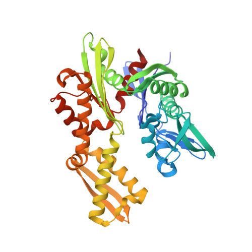
4IO8 - PubMed Abstract:
The molecular chaperones of the Hsp70 family have been recognized as targets for anti-cancer therapy. Since several paralogs of Hsp70 proteins exist in cytosol, endoplasmic reticulum and mitochondria, we investigated which isoform needs to be down-regulated for reducing viability of cancer cells. For two recently identified small molecule inhibitors, VER-155008 and 2-phenylethynesulfonamide (PES), which are proposed to target different sites in Hsp70s, we analyzed the molecular mode of action in vitro. We found that for significant reduction of viability of cancer cells simultaneous knockdown of heat-inducible Hsp70 (HSPA1) and constitutive Hsc70 (HSPA8) is necessary. The compound VER-155008, which binds to the nucleotide binding site of Hsp70, arrests the nucleotide binding domain (NBD) in a half-open conformation and thereby acts as ATP-competitive inhibitor that prevents allosteric control between NBD and substrate binding domain (SBD). Compound PES interacts with the SBD of Hsp70 in an unspecific, detergent-like fashion, under the conditions tested. None of the two inhibitors investigated was isoform-specific.
- Zentrum für Molekulare Biologie der Universität Heidelberg (ZMBH), DKFZ-ZMBH-Alliance, Heidelberg, Germany.
Organizational Affiliation: