4INX | pdb_00004inx

Structure of Pheromone-binding protein 1 in complex with (Z,Z)-11,13- hexadecadienol


Experimental Data Snapshot

  • Method: X-RAY DIFFRACTION
  • Resolution: 1.85 Å
  • R-Value Free: 
    0.214 (Depositor), 0.218 (DCC) 
  • R-Value Work: 
    0.168 (Depositor), 0.173 (DCC) 
  • R-Value Observed: 
    0.170 (Depositor) 

wwPDB Validation 3D Report Full Report

Validation slider image for 4INX

Ligand Structure Quality Assessment 


This is version 1.1 of the entry. See complete history

Literature

Crystallographic Observation of pH-Induced Conformational Changes in the Amyelois transitella Pheromone-Binding Protein AtraPBP1.

di Luccio, E.Ishida, Y.Leal, W.S.Wilson, D.K.

(2013) PLoS One 8: e53840-e53840

  • DOI: https://doi.org/10.1371/journal.pone.0053840
  • Primary Citation Related Structures: 
    4INW, 4INX

  • PubMed Abstract: 

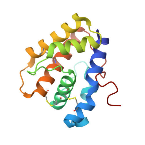
    The navel orangeworm, Amyelois transitella is a major agricultural pest causing large losses in a variety of tree crops. Control of this insect pest may be achieved by interfering with olfactory pathways to block detection of female-produced sex pheromones and consequently, disrupt mating. The first component of this pathway is the pheromone-binding protein AtraPBP1, which recognizes the pheromone and presents it to the odorant receptor housed in a sensory neuron of the male antennae. Release of the ligand depends on a pH-induced conformational change associated with the acidity of the membrane surface. To characterize this conformational change and to understand how pheromones bind, we have determined the high resolution crystal structures of AtraPBP1 in complex with two main constituents of the sex pheromone, i.e., (11Z,13Z)-hexadecadienal and (11Z,13Z)-hexadecadienol. Comparison with the structure of the unliganded form demonstrates a large ∼90° movement of the C-terminal helix which is observed in other pheromone- or odorant-binding proteins accompanied by an unpredicted 37° displacement of the N-terminal helix. Molecular dynamic trajectories suggest that the conformational change of the α1 helix facilitates the movement of the C-terminal helix.


  • Organizational Affiliation
    • Department of Molecular and Cellular Biology, University of California Davis, Davis, California, United States of America.

Macromolecule Content 

  • Total Structure Weight: 16.11 kDa 
  • Atom Count: 1,286 
  • Modeled Residue Count: 140 
  • Deposited Residue Count: 140 
  • Unique protein chains: 1

Macromolecules

Find similar proteins by:|  3D Structure
Entity ID: 1
MoleculeChains  Sequence LengthOrganismDetailsImage
Pheromone-binding protein 1140Amyelois transitellaMutation(s): 0 
UniProt
Find proteins for D0E9M1 (Amyelois transitella)
Explore D0E9M1 
Go to UniProtKB:  D0E9M1
Entity Groups
Sequence Clusters30% Identity50% Identity70% Identity90% Identity95% Identity100% Identity
UniProt GroupD0E9M1
Sequence Annotations
Expand
Reference Sequence

Small Molecules

Ligands 1 Unique
IDChains Name / Formula / InChI Key2D Diagram3D Interactions
1EX

Query on 1EX


Download:Ideal Coordinates CCD File
B [auth A](11Z,13Z)-hexadeca-11,13-dien-1-ol
C16 H30 O
GKFQVSXEEVMHMA-OUPQRBNQSA-N

Experimental Data & Validation

Experimental Data

  • Method: X-RAY DIFFRACTION
  • Resolution: 1.85 Å
  • R-Value Free:  0.214 (Depositor), 0.218 (DCC) 
  • R-Value Work:  0.168 (Depositor), 0.173 (DCC) 
  • R-Value Observed: 0.170 (Depositor) 
Space Group: P 65
Unit Cell:
Length ( Å )Angle ( ˚ )
a = 57.532α = 90
b = 57.532β = 90
c = 93.501γ = 120
Software Package:
Software NamePurpose
DENZOdata reduction
SCALEPACKdata scaling
PHASERphasing
REFMACrefinement
PDB_EXTRACTdata extraction
HKL-2000data collection

Structure Validation

View Full Validation Report


Ligand Structure Quality Assessment 


Entry History 

Deposition Data

Revision History  (Full details and data files)

  • Version 1.0: 2013-03-06
    Type: Initial release
  • Version 1.1: 2024-10-30
    Changes: Data collection, Database references, Derived calculations, Structure summary