4ILU

Crystal structure of Mycobacterium tuberculosis CarD


Experimental Data Snapshot

  • Method: X-RAY DIFFRACTION
  • Resolution: 2.30 Å
  • R-Value Free: 0.233 
  • R-Value Work: 0.207 
  • R-Value Observed: 0.208 

wwPDB Validation   3D Report Full Report


This is version 1.1 of the entry. See complete history


Literature

Crystal structure of Mycobacterium tuberculosis CarD, an essential RNA polymerase binding protein, reveals a quasidomain-swapped dimeric structural architecture.

Kaur, G.Dutta, D.Thakur, K.G.

(2014) Proteins 82: 879-884

  • DOI: https://doi.org/10.1002/prot.24419
  • Primary Citation of Related Structures:  
    4ILU, 4MFR

  • PubMed Abstract: 

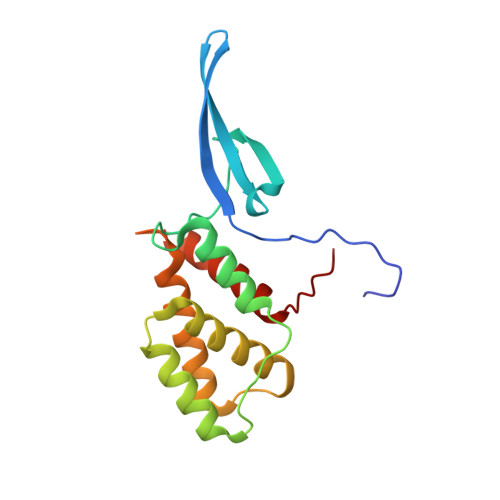
    Mycobacterium tuberculosis (Mtb) CarD is an essential transcriptional regulator that binds RNA polymerase and plays an important role in reprogramming transcription machinery under diverse stress conditions. Here, we report the crystal structure of CarD at 2.3 Å resolution, that represents the first structural description of CarD/CdnL-Like family of proteins. CarD adopts an overall bi-lobed structural architecture where N-terminal domain resembles 'tudor-like' domain and C-terminal domain adopts a novel five helical fold that lacks the predicted leucine zipper structural motif. The structure reveals dimeric state of CarD resulting from β-strand swapping between the N-terminal domains of each individual subunits. The structure provides crucial insights into the possible mode(s) of CarD/RNAP interactions.


  • Organizational Affiliation

    Structural Biology Laboratory, G. N. Ramachandran Protein Centre, CSIR-Institute of Microbial Technology, Chandigarh, 160036, India.


Macromolecules
Find similar proteins by:  (by identity cutoff)  |  3D Structure
Entity ID: 1
MoleculeChains Sequence LengthOrganismDetailsImage
RNA polymerase-binding transcription factor CarD170Mycobacterium tuberculosisMutation(s): 1 
Gene Names: carDMT3689Rv3583c
UniProt
Find proteins for P9WJG3 (Mycobacterium tuberculosis (strain ATCC 25618 / H37Rv))
Explore P9WJG3 
Go to UniProtKB:  P9WJG3
Entity Groups  
Sequence Clusters30% Identity50% Identity70% Identity90% Identity95% Identity100% Identity
UniProt GroupP9WJG3
Sequence Annotations
Expand
  • Reference Sequence
Experimental Data & Validation

Experimental Data

  • Method: X-RAY DIFFRACTION
  • Resolution: 2.30 Å
  • R-Value Free: 0.233 
  • R-Value Work: 0.207 
  • R-Value Observed: 0.208 
  • Space Group: P 41 21 2
Unit Cell:
Length ( Å )Angle ( ˚ )
a = 93.5α = 90
b = 93.5β = 90
c = 86.51γ = 90
Software Package:
Software NamePurpose
MOSFLMdata reduction
SCALAdata scaling
PHENIXrefinement
PDB_EXTRACTdata extraction
SOLVEphasing

Structure Validation

View Full Validation Report


Entry History 

Deposition Data

Revision History  (Full details and data files)

  • Version 1.0: 2013-10-30
    Type: Initial release
  • Version 1.1: 2022-08-24
    Changes: Database references, Derived calculations