The Mode of Inhibitor Binding to Peptidyl-tRNA Hydrolase: Binding Studies and Structure Determination of Unbound and Bound Peptidyl-tRNA Hydrolase from Acinetobacter baumannii
Kaushik, S., Singh, N., Yamini, S., Singh, A., Sinha, M., Arora, A., Kaur, P., Sharma, S., Singh, T.P.(2013) PLoS One 8: e67547-e67547
- PubMed: 23844024 Search on PubMedSearch on PubMed Central
- DOI: https://doi.org/10.1371/journal.pone.0067547
- Primary Citation Related Structures:
4FOP, 4FOT, 4HOY, 4IKO, 4JWK, 4JX9, 4JY7 - PubMed Abstract:
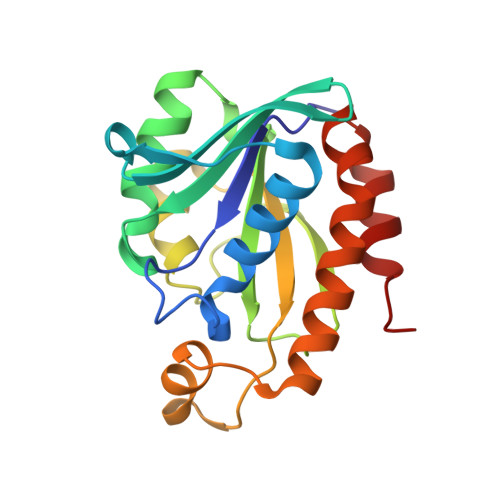
The incidences of infections caused by an aerobic Gram-negative bacterium, Acinetobacter baumannii are very common in hospital environments. It usually causes soft tissue infections including urinary tract infections and pneumonia. It is difficult to treat due to acquired resistance to available antibiotics is well known. In order to design specific inhibitors against one of the important enzymes, peptidyl-tRNA hydrolase from Acinetobacter baumannii, we have determined its three-dimensional structure. Peptidyl-tRNA hydrolase (AbPth) is involved in recycling of peptidyl-tRNAs which are produced in the cell as a result of premature termination of translation process. We have also determined the structures of two complexes of AbPth with cytidine and uridine. AbPth was cloned, expressed and crystallized in unbound and in two bound states with cytidine and uridine. The binding studies carried out using fluorescence spectroscopic and surface plasmon resonance techniques revealed that both cytidine and uridine bound to AbPth at nanomolar concentrations. The structure determinations of the complexes revealed that both ligands were located in the active site cleft of AbPth. The introduction of ligands to AbPth caused a significant widening of the entrance gate to the active site region and in the process of binding, it expelled several water molecules from the active site. As a result of interactions with protein atoms, the ligands caused conformational changes in several residues to attain the induced tight fittings. Such a binding capability of this protein makes it a versatile molecule for hydrolysis of peptidyl-tRNAs having variable peptide sequences. These are the first studies that revealed the mode of inhibitor binding in Peptidyl-tRNA hydrolases which will facilitate the structure based ligand design.
- Department of Biophysics, All India Institute of Medical Sciences, New Delhi, India.
Organizational Affiliation: