


Crystal structure of the complex of SETD8 with SAM
Yu, W., Tempel, W., Li, Y., El Bakkouri, M., Shapira, M., Bountra, C., Arrowsmith, C.H., Edwards, A.M., Brown, P.J., Structural Genomics Consortium (SGC)To be published.
Experimental Data Snapshot
Starting Model: experimental
View more details
Entity ID: 1 | |||||
|---|---|---|---|---|---|
| Molecule | Chains | Sequence Length | Organism | Details | Image |
| N-lysine methyltransferase SETD8 | 165 | Homo sapiens | Mutation(s): 1 Gene Names: SETD8, KMT5A, PRSET7, SET07, SET8 EC: 2.1.1 (PDB Primary Data), 2.1.1.43 (PDB Primary Data), 2.1.1.361 (UniProt) | 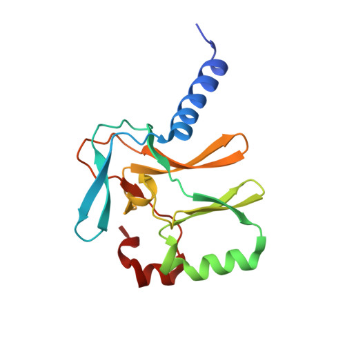 | |
UniProt & NIH Common Fund Data Resources | |||||
PHAROS: Q9NQR1 GTEx: ENSG00000183955 | |||||
Entity Groups | |||||
| Sequence Clusters | 30% Identity50% Identity70% Identity90% Identity95% Identity100% Identity | ||||
| UniProt Group | Q9NQR1 | ||||
Sequence AnnotationsExpand | |||||
Reference Sequence | |||||
Entity ID: 2 | |||||
|---|---|---|---|---|---|
| Molecule | Chains | Sequence Length | Organism | Details | Image |
| helical peptide | C [auth I] | 10 | unidentified | Mutation(s): 0 | 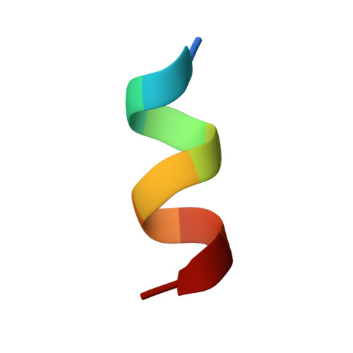 |
| Ligands 2 Unique | |||||
|---|---|---|---|---|---|
| ID | Chains | Name / Formula / InChI Key | 2D Diagram | 3D Interactions | |
| SAM Download:Ideal Coordinates CCD File | D [auth A], R [auth B] | S-ADENOSYLMETHIONINE C15 H22 N6 O5 S MEFKEPWMEQBLKI-FCKMPRQPSA-N |  | ||
| UNX Download:Ideal Coordinates CCD File | AA [auth B] E [auth A] F [auth A] G [auth A] H [auth A] | UNKNOWN ATOM OR ION X |  | ||
| Length ( Å ) | Angle ( ˚ ) |
|---|---|
| a = 101.439 | α = 90 |
| b = 101.439 | β = 90 |
| c = 140.799 | γ = 120 |
| Software Name | Purpose |
|---|---|
| SCALA | data scaling |
| PHASER | phasing |
| REFMAC | refinement |
| PDB_EXTRACT | data extraction |
| XDS | data reduction |


