4II9 | pdb_00004ii9

Crystal structure of Weissella viridescens FemXVv non-ribosomal amino acid transferase in complex with a peptidyl-RNA conjugate


Experimental Data Snapshot

  • Method: X-RAY DIFFRACTION
  • Resolution: 1.66 Å
  • R-Value Free: 
    0.209 (Depositor), 0.201 (DCC) 
  • R-Value Work: 
    0.175 (Depositor), 0.167 (DCC) 
  • R-Value Observed: 
    0.177 (Depositor) 

Starting Model: experimental
View more details

wwPDB Validation 3D Report Full Report

Validation slider image for 4II9

Ligand Structure Quality Assessment 


This is version 2.2 of the entry. See complete history

Literature

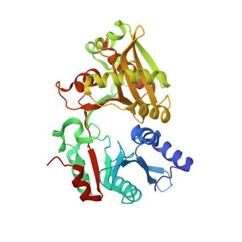
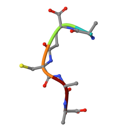
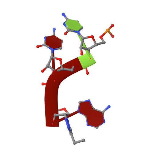
The Structure of FemXWv in Complex with a Peptidyl-RNA Conjugate: Mechanism of Aminoacyl Transfer from Ala-tRNA(Ala) to Peptidoglycan Precursors

Fonvielle, M.Li de La Sierra-Gallay, I.El-Sagheer, A.H.Lecerf, M.Patin, D.Mellal, D.Mayer, C.Blanot, D.Gale, N.Brown, T.van Tilbeurgh, H.Etheve-Quelquejeu, M.Arthur, M.

(2013) Angew Chem Int Ed Engl 52: 7278-7281

Macromolecule Content 

  • Total Structure Weight: 41.44 kDa 
  • Atom Count: 3,149 
  • Modeled Residue Count: 345 
  • Deposited Residue Count: 351 
  • Unique protein chains: 2
  • Unique nucleic acid chains: 1

Macromolecules


Find similar proteins by:|  3D Structure
Entity ID: 1
MoleculeChains  Sequence LengthOrganismDetailsImage
FemX343Weissella viridescensMutation(s): 0 
Gene Names: femX
EC: 2.3.2.10
UniProt
Find proteins for Q9EY50 (Weissella viridescens)
Explore Q9EY50 
Go to UniProtKB:  Q9EY50
Entity Groups
Sequence Clusters30% Identity50% Identity70% Identity90% Identity95% Identity100% Identity
UniProt GroupQ9EY50
Sequence Annotations
Expand
Reference Sequence
Find similar proteins by:  Sequence   |   3D Structure  
Entity ID: 2
MoleculeChains  Sequence LengthOrganismDetailsImage
5-mer peptide5N/AMutation(s): 0 
Find similar nucleic acids by:  Sequence
Entity ID: 3
MoleculeChains LengthOrganismImage
RNA (5'-R(P*CP*CP*(A9Z))-3')3N/A
Sequence Annotations
Expand
Reference Sequence

Experimental Data & Validation

Experimental Data

  • Method: X-RAY DIFFRACTION
  • Resolution: 1.66 Å
  • R-Value Free:  0.209 (Depositor), 0.201 (DCC) 
  • R-Value Work:  0.175 (Depositor), 0.167 (DCC) 
  • R-Value Observed: 0.177 (Depositor) 
Space Group: P 1 21 1
Unit Cell:
Length ( Å )Angle ( ˚ )
a = 42.13α = 90
b = 101.77β = 102.6
c = 46.32γ = 90
Software Package:
Software NamePurpose
MXdata collection
PHASERphasing
REFMACrefinement
XDSdata reduction
XSCALEdata scaling

Structure Validation

View Full Validation Report


Ligand Structure Quality Assessment 


Entry History 

Deposition Data

Revision History  (Full details and data files)

  • Version 1.0: 2013-07-03
    Type: Initial release
  • Version 1.1: 2013-08-07
    Changes: Database references
  • Version 2.0: 2020-07-29
    Type: Remediation
    Reason: Carbohydrate remediation
    Changes: Advisory, Atomic model, Data collection, Database references, Derived calculations, Structure summary
  • Version 2.1: 2023-11-08
    Changes: Data collection, Database references, Refinement description, Structure summary
  • Version 2.2: 2024-10-09
    Changes: Structure summary