4IG9

Structure of NAD-dependent protein deacetylase sirtuin-1 (open state, 2.64 A)


Experimental Data Snapshot

  • Method: X-RAY DIFFRACTION
  • Resolution: 2.64 Å
  • R-Value Free: 0.263 
  • R-Value Work: 0.227 
  • R-Value Observed: 0.228 

wwPDB Validation   3D Report Full Report


This is version 1.2 of the entry. See complete history


Literature

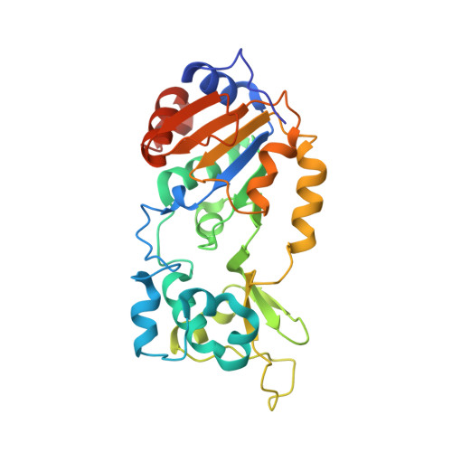
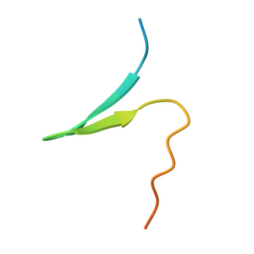
Structural and Functional Analysis of Human SIRT1.

Davenport, A.M.Huber, F.M.Hoelz, A.

(2014) J Mol Biol 426: 526-541

  • DOI: https://doi.org/10.1016/j.jmb.2013.10.009
  • Primary Citation of Related Structures:  
    4IG9, 4KXQ

  • PubMed Abstract: 

    SIRT1 is a NAD(+)-dependent deacetylase that plays important roles in many cellular processes. SIRT1 activity is uniquely controlled by a C-terminal regulatory segment (CTR). Here we present crystal structures of the catalytic domain of human SIRT1 in complex with the CTR in an open apo form and a closed conformation in complex with a cofactor and a pseudo-substrate peptide. The catalytic domain adopts the canonical sirtuin fold. The CTR forms a β hairpin structure that complements the β sheet of the NAD(+)-binding domain, covering an essentially invariant hydrophobic surface. The apo form adopts a distinct open conformation, in which the smaller subdomain of SIRT1 undergoes a rotation with respect to the larger NAD(+)-binding subdomain. A biochemical analysis identifies key residues in the active site, an inhibitory role for the CTR, and distinct structural features of the CTR that mediate binding and inhibition of the SIRT1 catalytic domain.


  • Organizational Affiliation

    Division of Chemistry and Chemical Engineering, California Institute of Technology, 1200 East California Boulevard, Pasadena, CA 91125, USA.


Macromolecules
Find similar proteins by:  (by identity cutoff)  |  3D Structure
Entity ID: 1
MoleculeChains Sequence LengthOrganismDetailsImage
NAD-dependent protein deacetylase sirtuin-1
A, C, E, G
281Homo sapiensMutation(s): 0 
Gene Names: SIR2L1SIRT1
EC: 3.5.1
UniProt & NIH Common Fund Data Resources
Find proteins for Q96EB6 (Homo sapiens)
Explore Q96EB6 
Go to UniProtKB:  Q96EB6
PHAROS:  Q96EB6
GTEx:  ENSG00000096717 
Entity Groups  
Sequence Clusters30% Identity50% Identity70% Identity90% Identity95% Identity100% Identity
UniProt GroupQ96EB6
Sequence Annotations
Expand
  • Reference Sequence
Find similar proteins by:  (by identity cutoff)  |  3D Structure
Entity ID: 2
MoleculeChains Sequence LengthOrganismDetailsImage
NAD-dependent protein deacetylase sirtuin-1
B, D, F, H
31Homo sapiensMutation(s): 0 
Gene Names: SIR2L1SIRT1
UniProt & NIH Common Fund Data Resources
Find proteins for Q96EB6 (Homo sapiens)
Explore Q96EB6 
Go to UniProtKB:  Q96EB6
PHAROS:  Q96EB6
GTEx:  ENSG00000096717 
Entity Groups  
Sequence Clusters30% Identity50% Identity70% Identity90% Identity95% Identity100% Identity
UniProt GroupQ96EB6
Sequence Annotations
Expand
  • Reference Sequence
Experimental Data & Validation

Experimental Data

  • Method: X-RAY DIFFRACTION
  • Resolution: 2.64 Å
  • R-Value Free: 0.263 
  • R-Value Work: 0.227 
  • R-Value Observed: 0.228 
  • Space Group: P 43 21 2
Unit Cell:
Length ( Å )Angle ( ˚ )
a = 115.781α = 90
b = 115.781β = 90
c = 350.511γ = 90
Software Package:
Software NamePurpose
SCALEPACKdata scaling
PHENIXrefinement
PDB_EXTRACTdata extraction
HKL-2000data collection
HKL-2000data reduction
SHARPphasing

Structure Validation

View Full Validation Report


Entry History 

Deposition Data

Revision History  (Full details and data files)

  • Version 1.0: 2013-11-06
    Type: Initial release
  • Version 1.1: 2014-02-05
    Changes: Database references
  • Version 1.2: 2024-02-28
    Changes: Data collection, Database references, Derived calculations