Cysteine Dioxygenase Structures from pH4 to 9: Consistent Cys-Persulfenate Formation at Intermediate pH and a Cys-Bound Enzyme at Higher pH.
Driggers, C.M., Cooley, R.B., Sankaran, B., Hirschberger, L.L., Stipanuk, M.H., Karplus, P.A.(2013) J Mol Biology 425: 3121-3136
- PubMed: 23747973 Search on PubMedSearch on PubMed Central
- DOI: https://doi.org/10.1016/j.jmb.2013.05.028
- Primary Citation Related Structures:
4IEO, 4IEP, 4IEQ, 4IER, 4IES, 4IET, 4IEU, 4IEV, 4IEW, 4IEX, 4IEY, 4IEZ, 4JTN, 4JTO - PubMed Abstract:
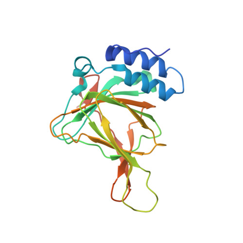
Mammalian cysteine dioxygenase (CDO) is a mononuclear non-heme iron protein that catalyzes the conversion of cysteine (Cys) to cysteine sulfinic acid by an unclarified mechanism. One structural study revealed that a Cys-persulfenate (or Cys-persulfenic acid) formed in the active site, but quantum mechanical calculations have been used to support arguments that it is not an energetically feasible reaction intermediate. Here, we report a series of high-resolution structures of CDO soaked with Cys at pH values from 4 to 9. Cys binding is minimal at pH≤5 and persulfenate formation is consistently seen at pH values between 5.5 and 7. Also, a structure determined using laboratory-based X-ray diffraction shows that the persulfenate, with an apparent average O-O separation distance of ~1.8Å, is not an artifact of synchrotron radiation. At pH≥8, the active-site iron shifts from 4- to 5-coordinate, and Cys soaks reveal a complex with Cys, but no dioxygen, bound. This 'Cys-only' complex differs in detail from a previously published 'Cys-only' complex, which we reevaluate and conclude is not reliable. The high-resolution structures presented here do not resolve the CDO mechanism but do imply that an iron-bound persulfenate (or persulfenic acid) is energetically accessible in the CDO active site, and that CDO active-site chemistry in the crystals is influenced by protonation/deprotonation events with effective pKa values near ~5.5 and ~7.5 that influence Cys binding and oxygen binding/reactivity, respectively. Furthermore, this work provides reliable ligand-bound models for guiding future mechanistic considerations.
- Department of Biochemistry and Biophysics, 2011 Ag and Life Sciences Building, Oregon State University, Corvallis, OR 97331, USA.
Organizational Affiliation: