4IDZ | pdb_00004idz

Crystal structure of the human fat mass and obesity associated protein (FTO) in complex with N-oxalylglycine (NOG)


Experimental Data Snapshot

  • Method: X-RAY DIFFRACTION
  • Resolution: 2.46 Å
  • R-Value Free: 
    0.245 (Depositor), 0.202 (DCC) 
  • R-Value Work: 
    0.194 (Depositor), 0.207 (DCC) 

Starting Model: experimental
View more details

wwPDB Validation 3D Report Full Report

Validation slider image for 4IDZ

This is version 1.5 of the entry. See complete history

Literature

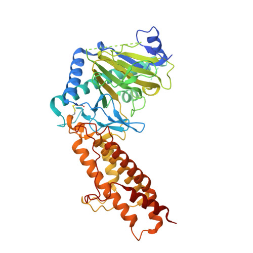
Structural basis for inhibition of the fat mass and obesity associated protein (FTO)

Aik, W.S.Demetriades, M.Hamdan, M.K.K.Bagg, E.A.L.Yeoh, K.K.Lejeune, C.Zhang, Z.McDonough, M.A.Schofield, C.J.

(2013) J Med Chem 56: 3680-3688

  • DOI: https://doi.org/10.1021/jm400193d
  • Primary Citation Related Structures: 
    4IDZ, 4IE0, 4IE4, 4IE5, 4IE6, 4IE7

  • PubMed Abstract: 

    The fat mass and obesity associated protein (FTO) is a potential target for anti-obesity medicines. FTO is a 2-oxoglutarate (2OG)-dependent N-methyl nucleic acid demethylase that acts on substrates including 3-methylthymidine, 3-methyluracil, and 6-methyladenine. To identify FTO inhibitors, we screened a set of 2OG analogues and related compounds using differential scanning fluorometry- and liquid chromatography-based assays. The results revealed sets of both cyclic and acyclic 2OG analogues that are FTO inhibitors. Identified inhibitors include small molecules that have been used in clinical studies for the inhibition of other 2OG oxygenases. Crystallographic analyses reveal inhibition by 2OG cosubstrate or primary substrate competitors as well as compounds that bind across both cosubstrate and primary substrate binding sites. The results will aid the development of more potent and selective FTO inhibitors.


  • Organizational Affiliation
    • Chemistry Research Laboratory, University of Oxford , 12 Mansfield Road, Oxford OX1 3TA, United Kingdom.

Macromolecule Content 

  • Total Structure Weight: 57.12 kDa 
  • Atom Count: 3,554 
  • Modeled Residue Count: 437 
  • Deposited Residue Count: 495 
  • Unique protein chains: 1

Macromolecules

Find similar proteins by:|  3D Structure
Entity ID: 1
MoleculeChains  Sequence LengthOrganismDetailsImage
Alpha-ketoglutarate-dependent dioxygenase FTO495Homo sapiensMutation(s): 0 
Gene Names: FTOKIAA1752
EC: 1.14.11 (PDB Primary Data), 1.14.11.53 (UniProt)
UniProt & NIH Common Fund Data Resources
Find proteins for Q9C0B1 (Homo sapiens)
Explore Q9C0B1 
Go to UniProtKB:  Q9C0B1
PHAROS:  Q9C0B1
GTEx:  ENSG00000140718 
Entity Groups
Sequence Clusters30% Identity50% Identity70% Identity90% Identity95% Identity100% Identity
UniProt GroupQ9C0B1
Sequence Annotations
Expand
Reference Sequence

Small Molecules

Binding Affinity Annotations 
IDSourceBinding Affinity
OGA BindingDB:  4IDZ IC50: min: 3.65e+4, max: 4.40e+4 (nM) from 2 assay(s)

Experimental Data & Validation

Experimental Data

  • Method: X-RAY DIFFRACTION
  • Resolution: 2.46 Å
  • R-Value Free:  0.245 (Depositor), 0.202 (DCC) 
  • R-Value Work:  0.194 (Depositor), 0.207 (DCC) 
Space Group: H 3
Unit Cell:
Length ( Å )Angle ( ˚ )
a = 140.412α = 90
b = 140.412β = 90
c = 83.447γ = 120
Software Package:
Software NamePurpose
DENZOdata reduction
SCALEPACKdata scaling
PHASERphasing
PHENIXrefinement
PDB_EXTRACTdata extraction
CrystalCleardata collection
HKL-2000data reduction
HKL-2000data scaling

Structure Validation

View Full Validation Report


Entry History 

Deposition Data

Revision History  (Full details and data files)

  • Version 1.0: 2013-04-03
    Type: Initial release
  • Version 1.1: 2013-04-17
    Changes: Database references
  • Version 1.2: 2013-07-31
    Changes: Database references
  • Version 1.3: 2017-11-15
    Changes: Refinement description
  • Version 1.4: 2023-11-08
    Changes: Data collection, Database references, Derived calculations, Refinement description
  • Version 1.5: 2024-11-20
    Changes: Structure summary