4ICU | pdb_00004icu

Ubiquitin-like domain of human tubulin folding cofactor E - crystal from A


Experimental Data Snapshot

  • Method: X-RAY DIFFRACTION
  • Resolution: 2.40 Å
  • R-Value Free: 
    0.263 (Depositor) 
  • R-Value Work: 
    0.204 (Depositor) 
  • R-Value Observed: 
    0.207 (Depositor) 

Starting Model: experimental
View more details

wwPDB Validation 3D Report Full Report

Validation slider image for 4ICU

This is version 1.2 of the entry. See complete history

Literature

The structure of the complex between alpha-tubulin, TBCE and TBCB reveals a tubulin dimer dissociation mechanism.

Serna, M.Carranza, G.Martin-Benito, J.Janowski, R.Canals, A.Coll, M.Zabala, J.C.Valpuesta, J.M.

(2015) J Cell Sci 128: 1824-1834

  • DOI: https://doi.org/10.1242/jcs.167387
  • Primary Citation Related Structures: 
    4ICU, 4ICV

  • PubMed Abstract: 

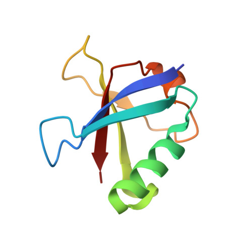
    Tubulin proteostasis is regulated by a group of molecular chaperones termed tubulin cofactors (TBC). Whereas tubulin heterodimer formation is well-characterized biochemically, its dissociation pathway is not clearly understood. Here, we carried out biochemical assays to dissect the role of the human TBCE and TBCB chaperones in α-tubulin-β-tubulin dissociation. We used electron microscopy and image processing to determine the three-dimensional structure of the human TBCE, TBCB and α-tubulin (αEB) complex, which is formed upon α-tubulin-β-tubulin heterodimer dissociation by the two chaperones. Docking the atomic structures of domains of these proteins, including the TBCE UBL domain, as we determined by X-ray crystallography, allowed description of the molecular architecture of the αEB complex. We found that heterodimer dissociation is an energy-independent process that takes place through a disruption of the α-tubulin-β-tubulin interface that is caused by a steric interaction between β-tubulin and the TBCE cytoskeleton-associated protein glycine-rich (CAP-Gly) and leucine-rich repeat (LRR) domains. The protruding arrangement of chaperone ubiquitin-like (UBL) domains in the αEB complex suggests that there is a direct interaction of this complex with the proteasome, thus mediating α-tubulin degradation.


  • Organizational Affiliation
    • Departamento de Estructura de Macromoléculas, Centro Nacional de Biotecnología (CNB-CSIC), Madrid 28049, Spain.

Macromolecule Content 

  • Total Structure Weight: 39.28 kDa 
  • Atom Count: 2,838 
  • Modeled Residue Count: 336 
  • Deposited Residue Count: 340 
  • Unique protein chains: 1

Macromolecules

Find similar proteins by:|  3D Structure
Entity ID: 1
MoleculeChains  Sequence LengthOrganismDetailsImage
Tubulin-specific chaperone E
A, B, C, D
85Homo sapiensMutation(s): 0 
Gene Names: TBCE
UniProt & NIH Common Fund Data Resources
Find proteins for Q15813 (Homo sapiens)
Explore Q15813 
Go to UniProtKB:  Q15813
PHAROS:  Q15813
Entity Groups
Sequence Clusters30% Identity50% Identity70% Identity90% Identity95% Identity100% Identity
UniProt GroupQ15813
Sequence Annotations
Expand
Reference Sequence

Experimental Data & Validation

Experimental Data

  • Method: X-RAY DIFFRACTION
  • Resolution: 2.40 Å
  • R-Value Free:  0.263 (Depositor) 
  • R-Value Work:  0.204 (Depositor) 
  • R-Value Observed: 0.207 (Depositor) 
Space Group: P 21 21 21
Unit Cell:
Length ( Å )Angle ( ˚ )
a = 43.2α = 90
b = 60.11β = 90
c = 130.45γ = 90
Software Package:
Software NamePurpose
DNAdata collection
PHASERphasing
REFMACrefinement
XDSdata reduction
SCALAdata scaling

Structure Validation

View Full Validation Report


Entry History 

Deposition Data

Revision History  (Full details and data files)

  • Version 1.0: 2014-06-18
    Type: Initial release
  • Version 1.1: 2016-06-22
    Changes: Database references
  • Version 1.2: 2023-09-20
    Changes: Data collection, Database references, Refinement description