4IC5 | pdb_00004ic5

Crystal structure of Deg5


Experimental Data Snapshot

  • Method: X-RAY DIFFRACTION
  • Resolution: 2.61 Å
  • R-Value Free: 
    0.232 (Depositor), 0.229 (DCC) 
  • R-Value Work: 
    0.182 (Depositor), 0.179 (DCC) 
  • R-Value Observed: 
    0.184 (Depositor) 

Starting Model: experimental
View more details

wwPDB Validation 3D Report Full Report

Validation slider image for 4IC5

This is version 1.2 of the entry. See complete history

Literature

The structures of Arabidopsis Deg5 and Deg8 reveal new insights into HtrA proteases

Sun, W.Gao, F.Fan, H.Shan, X.Sun, R.Liu, L.Gong, W.

(2013) Acta Crystallogr D Biol Crystallogr 69: 830-837

  • DOI: https://doi.org/10.1107/S0907444913002023
  • Primary Citation Related Structures: 
    4IC5, 4IC6

  • PubMed Abstract: 

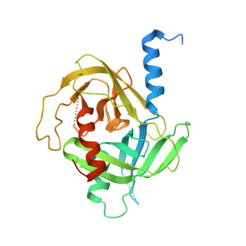
    Plant Deg5 and Deg8 are two members of the HtrA proteases, a family of oligomeric serine endopeptidases that are involved in a variety of protein quality-control processes. These two HtrA proteases are located in the thylakoid lumen and participate in high-light stress responses by collaborating with other chloroplast proteins. Deg5 and Deg8 degrade photodamaged D1 protein of the photosystem II reaction centre, allowing its in situ replacement. Here, the crystal structures of Arabidopsis thaliana Deg5 (S266A) and Deg8 (S292A) are reported at 2.6 and 2.0 Å resolution, respectively. The Deg5 trimer contains two calcium ions in a central channel, suggesting a link between photodamage control and calcium ions in chloroplasts. Previous structures of HtrA proteases have indicated that their regulation usually requires C-terminal PDZ domain(s). Deg5 is unique in that it contains no PDZ domain and the trimeric structure of Deg5 (S266A) reveals a novel catalytic triad conformation. A similar triad conformation is observed in the hexameric structure of the single PDZ-domain-containing Deg8 (S292A). These findings suggest a novel activation mechanism for plant HtrA proteases and provide structural clues to their function in light-stress response.


  • Organizational Affiliation
    • Laboratory of Non-coding RNA, Institute of Biophysics, Chinese Academy of Sciences, 5 Datun Road, Chaoyang District, Beijing 100101, People's Republic of China.

Macromolecule Content 

  • Total Structure Weight: 94.67 kDa 
  • Atom Count: 4,946 
  • Modeled Residue Count: 629 
  • Deposited Residue Count: 873 
  • Unique protein chains: 1

Macromolecules

Find similar proteins by:|  3D Structure
Entity ID: 1
MoleculeChains  Sequence LengthOrganismDetailsImage
Protease Do-like 5, chloroplastic
A, B, C
291Arabidopsis thalianaMutation(s): 1 
Gene Names: DEGP5HHOAAt4g18370F28J12.30
EC: 3.4.21
UniProt
Find proteins for Q9SEL7 (Arabidopsis thaliana)
Explore Q9SEL7 
Go to UniProtKB:  Q9SEL7
Entity Groups
Sequence Clusters30% Identity50% Identity70% Identity90% Identity95% Identity100% Identity
UniProt GroupQ9SEL7
Sequence Annotations
Expand
Reference Sequence

Experimental Data & Validation

Experimental Data

  • Method: X-RAY DIFFRACTION
  • Resolution: 2.61 Å
  • R-Value Free:  0.232 (Depositor), 0.229 (DCC) 
  • R-Value Work:  0.182 (Depositor), 0.179 (DCC) 
  • R-Value Observed: 0.184 (Depositor) 
Space Group: C 1 2 1
Unit Cell:
Length ( Å )Angle ( ˚ )
a = 109.09α = 90
b = 125.952β = 102.91
c = 83.325γ = 90
Software Package:
Software NamePurpose
HKL-2000data collection
PHASESphasing
PHENIXrefinement
HKL-2000data reduction
HKL-2000data scaling

Structure Validation

View Full Validation Report


Entry History 

Deposition Data

Revision History  (Full details and data files)

  • Version 1.0: 2013-05-01
    Type: Initial release
  • Version 1.1: 2013-06-26
    Changes: Database references
  • Version 1.2: 2023-11-08
    Changes: Data collection, Database references, Derived calculations, Refinement description