4I8N | pdb_00004i8n

CRYSTAL STRUCTURE of PROTEIN TYROSINE PHOSPHATASE 1B IN COMPLEX WITH AN INHIBITOR [(4-{(2S)-2-(1,3-BENZOXAZOL-2-YL)-2-[(4-FLUOROPHENYL)SULFAMOYL]ETHYL}PHENYL)AMINO](OXO)ACETIC ACID


Experimental Data Snapshot

  • Method: X-RAY DIFFRACTION
  • Resolution: 2.50 Å
  • R-Value Free: 
    0.248 (Depositor), 0.253 (DCC) 
  • R-Value Work: 
    0.184 (Depositor), 0.193 (DCC) 
  • R-Value Observed: 
    0.188 (Depositor) 

wwPDB Validation 3D Report Full Report

Validation slider image for 4I8N

Ligand Structure Quality Assessment 


This is version 1.2 of the entry. See complete history

Literature

X-Ray Structure of PTP1B in Complex with a New PTP1B Inhibitor.

Reddy, M.V.Ghadiyaram, C.Panigrahi, S.K.Krishnamurthy, N.R.Hosahalli, S.Chandrasekharappa, A.P.Manna, D.Badiger, S.E.Dubey, P.K.Mangamoori, L.N.

(2014) Protein Pept Lett 21: 90-93

  • DOI: https://doi.org/10.2174/09298665113209990089
  • Primary Citation Related Structures: 
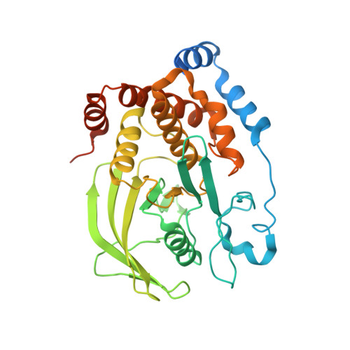
    4I8N

  • PubMed Abstract: 

    Protein tyrosine phosphatase 1B (PTP1B) is a prototype non receptor cytoplasmic PTPase enzyme that has been implicated in regulation of insulin and leptin signaling pathways. Studies on PTP1B knockout mice and PTP1B antisense treated mice suggested that inhibition of PTP1B would be an effective strategy for the treatment of type II diabetes and obesity. Here we report the X-ray structure of PTP1B in complex with compound IN1834-146C (PDB ID 4I8N). The crystals belong to P3121 space group with cell dimensions (a = b = 87.89 Å, c = 103.68 Å) diffracted to 2.5 Å. The crystal structure contained one molecule of protein in the asymmetric unit and was solved by molecular replacement method. The compound engages both catalytic site and allosteric sites of PTP1B protein. We described the molecular interaction of the compound with the active site residues of PTP1B in this crystal structure report.


  • Organizational Affiliation
    • Aurigene Discovery Technologies Ltd, 39-40 KIADB Industrial Area, Phase II, Electronic city, Hosur Road, Bangalore-560100, India. manusekhar1975@gmail.com.

Macromolecule Content 

  • Total Structure Weight: 41.29 kDa 
  • Atom Count: 2,526 
  • Modeled Residue Count: 298 
  • Deposited Residue Count: 354 
  • Unique protein chains: 1

Macromolecules

Find similar proteins by:|  3D Structure
Entity ID: 1
MoleculeChains  Sequence LengthOrganismDetailsImage
Tyrosine-protein phosphatase non-receptor type 1354Homo sapiensMutation(s): 0 
Gene Names: PTPN1PTP1B
EC: 3.1.3.48
UniProt & NIH Common Fund Data Resources
Find proteins for P18031 (Homo sapiens)
Explore P18031 
Go to UniProtKB:  P18031
PHAROS:  P18031
GTEx:  ENSG00000196396 
Entity Groups
Sequence Clusters30% Identity50% Identity70% Identity90% Identity95% Identity100% Identity
UniProt GroupP18031
Sequence Annotations
Expand
Reference Sequence

Small Molecules

Ligands 1 Unique
IDChains Name / Formula / InChI Key2D Diagram3D Interactions
1CG

Query on 1CG


Download:Ideal Coordinates CCD File
B [auth A][(4-{(2S)-2-(1,3-benzoxazol-2-yl)-2-[(4-fluorophenyl)sulfamoyl]ethyl}phenyl)amino](oxo)acetic acid
C23 H18 F N3 O6 S
ZSINWSQBKYZRCH-FQEVSTJZSA-N

Experimental Data & Validation

Experimental Data

  • Method: X-RAY DIFFRACTION
  • Resolution: 2.50 Å
  • R-Value Free:  0.248 (Depositor), 0.253 (DCC) 
  • R-Value Work:  0.184 (Depositor), 0.193 (DCC) 
  • R-Value Observed: 0.188 (Depositor) 
Space Group: P 31 2 1
Unit Cell:
Length ( Å )Angle ( ˚ )
a = 87.89α = 90
b = 87.89β = 90
c = 103.68γ = 120
Software Package:
Software NamePurpose
CrystalCleardata collection
AMoREphasing
REFMACrefinement
DENZOdata reduction
SCALEPACKdata scaling

Structure Validation

View Full Validation Report


Ligand Structure Quality Assessment 


Entry History 

Deposition Data

Revision History  (Full details and data files)

  • Version 1.0: 2012-12-26
    Type: Initial release
  • Version 1.1: 2014-01-01
    Changes: Database references
  • Version 1.2: 2024-02-28
    Changes: Data collection, Database references, Derived calculations