4I8M | pdb_00004i8m

Crystal structure of rabbit Ryanodine receptor 1 (residues 1-536) disease mutant V219I


Experimental Data Snapshot

  • Method: X-RAY DIFFRACTION
  • Resolution: 2.80 Å
  • R-Value Free: 
    0.253 (Depositor), 0.254 (DCC) 
  • R-Value Work: 
    0.235 (Depositor), 0.234 (DCC) 
  • R-Value Observed: 
    0.236 (Depositor) 

Starting Model: experimental
View more details

wwPDB Validation 3D Report Full Report

Validation slider image for 4I8M

This is version 1.2 of the entry. See complete history

Literature

Disease mutations in the ryanodine receptor N-terminal region couple to a mobile intersubunit interface.

Kimlicka, L.Lau, K.Tung, C.C.Van Petegem, F.

(2013) Nat Commun 4: 1506-1506

  • DOI: https://doi.org/10.1038/ncomms2501
  • Primary Citation Related Structures: 
    4I0Y, 4I1E, 4I2S, 4I37, 4I3N, 4I6I, 4I7I, 4I8M, 4I96

  • PubMed Abstract: 

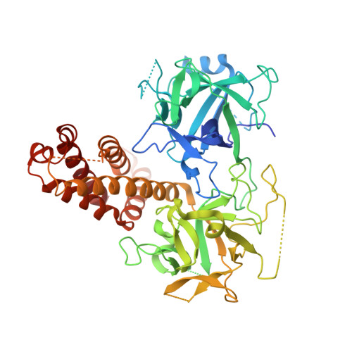
    Ryanodine receptors are large channels that release Ca(2+) from the endoplasmic and sarcoplasmic reticulum. Hundreds of RyR mutations can cause cardiac and skeletal muscle disorders, yet detailed mechanisms explaining their effects have been lacking. Here we compare pseudo-atomic models and propose that channel opening coincides with widening of a cytoplasmic vestibule formed by the N-terminal region, thus altering an interface targeted by 20 disease mutations. We solve crystal structures of several disease mutants that affect intrasubunit domain-domain interfaces. Mutations affecting intrasubunit ionic pairs alter relative domain orientations, and thus couple to surrounding interfaces. Buried disease mutations cause structural changes that also connect to the intersubunit contact area. These results suggest that the intersubunit contact region between N-terminal domains is a prime target for disease mutations, direct or indirect, and we present a model whereby ryanodine receptors and inositol-1,4,5-trisphosphate receptors are activated by altering domain arrangements in the N-terminal region.


  • Organizational Affiliation
    • Department of Biochemistry and Molecular Biology, Life Sciences Institute, University of British Columbia, Vancouver, British Columbia, Canada V6T 1Z3.

Macromolecule Content 

  • Total Structure Weight: 59.58 kDa 
  • Atom Count: 3,620 
  • Modeled Residue Count: 480 
  • Deposited Residue Count: 536 
  • Unique protein chains: 1

Macromolecules

Find similar proteins by:|  3D Structure
Entity ID: 1
MoleculeChains  Sequence LengthOrganismDetailsImage
Ryanodine receptor 1536Oryctolagus cuniculusMutation(s): 1 
Gene Names: RYR1
UniProt
Find proteins for P11716 (Oryctolagus cuniculus)
Explore P11716 
Go to UniProtKB:  P11716
Entity Groups
Sequence Clusters30% Identity50% Identity70% Identity90% Identity95% Identity100% Identity
UniProt GroupP11716
Sequence Annotations
Expand
Reference Sequence

Small Molecules

Ligands 1 Unique
IDChains Name / Formula / InChI Key2D Diagram3D Interactions
GOL

Query on GOL


Download:Ideal Coordinates CCD File
B [auth A]GLYCEROL
C3 H8 O3
PEDCQBHIVMGVHV-UHFFFAOYSA-N

Experimental Data & Validation

Experimental Data

  • Method: X-RAY DIFFRACTION
  • Resolution: 2.80 Å
  • R-Value Free:  0.253 (Depositor), 0.254 (DCC) 
  • R-Value Work:  0.235 (Depositor), 0.234 (DCC) 
  • R-Value Observed: 0.236 (Depositor) 
Space Group: H 3 2
Unit Cell:
Length ( Å )Angle ( ˚ )
a = 169.267α = 90
b = 169.267β = 90
c = 305.599γ = 120
Software Package:
Software NamePurpose
MxDCdata collection
PHASERphasing
REFMACrefinement
SCALAdata scaling

Structure Validation

View Full Validation Report


Entry History 

Deposition Data

Revision History  (Full details and data files)

  • Version 1.0: 2013-02-20
    Type: Initial release
  • Version 1.1: 2013-03-06
    Changes: Database references
  • Version 1.2: 2023-09-20
    Changes: Data collection, Database references, Derived calculations, Refinement description