4I7D

Siah1 bound to synthetic peptide (ACE)KLRPVAMVRP(PRK)VR


Experimental Data Snapshot

  • Method: X-RAY DIFFRACTION
  • Resolution: 2.40 Å
  • R-Value Free: 0.202 
  • R-Value Work: 0.171 
  • R-Value Observed: 0.173 

wwPDB Validation   3D Report Full Report


This is version 1.2 of the entry. See complete history


Literature

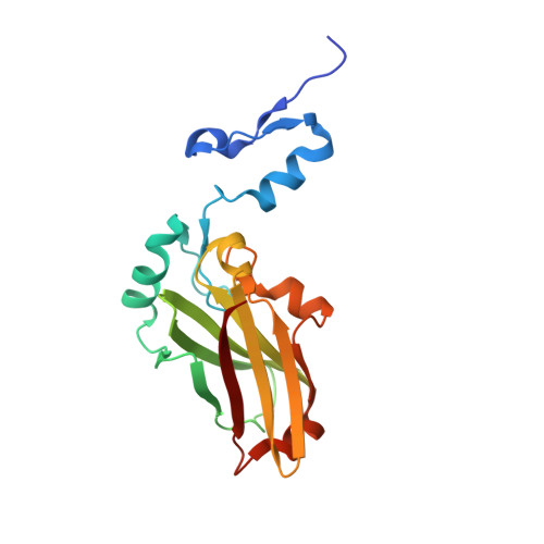
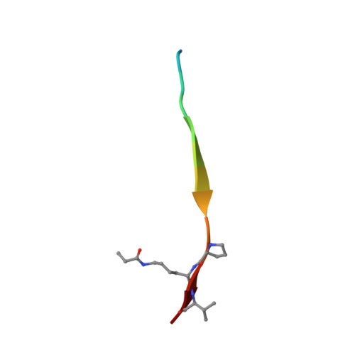
Structure-based design of covalent siah inhibitors.

Stebbins, J.L.Santelli, E.Feng, Y.De, S.K.Purves, A.Motamedchaboki, K.Wu, B.Ronai, Z.A.Liddington, R.C.Pellecchia, M.

(2013) Chem Biol 20: 973-982

  • DOI: https://doi.org/10.1016/j.chembiol.2013.06.008
  • Primary Citation of Related Structures:  
    4I7B, 4I7C, 4I7D

  • PubMed Abstract: 
  • The E3 ubiquitin ligase Siah regulates key cellular events that are central to cancer development and progression. A promising route to Siah inhibition is disrupting its interactions with adaptor proteins. However, typical of protein-protein interactions, traditional unbiased approaches to ligand discovery did not produce viable hits against this target, despite considerable effort and a multitude of approaches ...

    The E3 ubiquitin ligase Siah regulates key cellular events that are central to cancer development and progression. A promising route to Siah inhibition is disrupting its interactions with adaptor proteins. However, typical of protein-protein interactions, traditional unbiased approaches to ligand discovery did not produce viable hits against this target, despite considerable effort and a multitude of approaches. Ultimately, a rational structure-based design strategy was successful for the identification of Siah inhibitors in which peptide binding drives specific covalent bond formation with the target. X-ray crystallography, mass spectrometry, and functional data demonstrate that these peptide mimetics are efficient covalent inhibitors of Siah and antagonize Siah-dependent regulation of Erk and Hif signaling in the cell. The proposed strategy may result useful as a general approach to the design of peptide-based inhibitors of other protein-protein interactions.


    Organizational Affiliation

    Signal Transduction Program and Cell Death Program, Cancer Center, Sanford-Burnham Medical Research Institute, 10901 North Torrey Pines Road, La Jolla, CA 92037, USA.


Macromolecules
Find similar proteins by:  (by identity cutoff)  |  3D Structure
Entity ID: 1
MoleculeChainsSequence LengthOrganismDetailsImage
E3 ubiquitin-protein ligase SIAH1
A, C
196Homo sapiensMutation(s): 0 
Gene Names: HUMSIAHSIAH1
EC: 6.3.2 (PDB Primary Data), 2.3.2.27 (UniProt)
UniProt & NIH Common Fund Data Resources
Find proteins for Q8IUQ4 (Homo sapiens)
Explore Q8IUQ4 
Go to UniProtKB:  Q8IUQ4
PHAROS:  Q8IUQ4
Entity Groups  
Sequence Clusters30% Identity50% Identity70% Identity90% Identity95% Identity100% Identity
UniProt GroupQ8IUQ4
Protein Feature View
Expand
  • Reference Sequence

Find similar proteins by:  Sequence   |   3D Structure  

Entity ID: 2
MoleculeChainsSequence LengthOrganismDetailsImage
Protein phyllopod
B, D
14Drosophila melanogasterMutation(s): 2 
Gene Names: phylCG10108
UniProt
Find proteins for Q27934 (Drosophila melanogaster)
Explore Q27934 
Go to UniProtKB:  Q27934
Entity Groups  
Sequence Clusters30% Identity50% Identity70% Identity90% Identity95% Identity100% Identity
UniProt GroupQ27934
Protein Feature View
Expand
  • Reference Sequence
Small Molecules
Ligands 2 Unique
IDChainsName / Formula / InChI Key2D Diagram3D Interactions
MPD
Query on MPD

Download Ideal Coordinates CCD File 
G [auth A](4S)-2-METHYL-2,4-PENTANEDIOL
C6 H14 O2
SVTBMSDMJJWYQN-YFKPBYRVSA-N
 Ligand Interaction
ZN
Query on ZN

Download Ideal Coordinates CCD File 
E [auth A],
F [auth A],
H [auth C],
I [auth C]
ZINC ION
Zn
PTFCDOFLOPIGGS-UHFFFAOYSA-N
 Ligand Interaction
Modified Residues  1 Unique
IDChainsTypeFormula2D DiagramParent
PRK
Query on PRK
B, D
L-PEPTIDE LINKINGC9 H18 N2 O3LYS
Biologically Interesting Molecules (External Reference) 1 Unique
Experimental Data & Validation

Experimental Data

  • Method: X-RAY DIFFRACTION
  • Resolution: 2.40 Å
  • R-Value Free: 0.202 
  • R-Value Work: 0.171 
  • R-Value Observed: 0.173 
  • Space Group: I 2 3
Unit Cell:
Length ( Å )Angle ( ˚ )
a = 160.977α = 90
b = 160.977β = 90
c = 160.977γ = 90
Software Package:
Software NamePurpose
Blu-Icedata collection
PHASERphasing
REFMACrefinement
HKL-2000data reduction
HKL-2000data scaling

Structure Validation

View Full Validation Report


Entry History 

Deposition Data

Revision History  (Full details and data files)

  • Version 1.0: 2013-08-14
    Type: Initial release
  • Version 1.1: 2013-09-11
    Changes: Database references
  • Version 1.2: 2023-09-20
    Changes: Data collection, Database references, Derived calculations, Refinement description