4I5Z | pdb_00004i5z

Insulin protein crystallization via langmuir-blodgett


Experimental Data Snapshot

  • Method: X-RAY DIFFRACTION
  • Resolution: 1.80 Å
  • R-Value Free: 
    0.218 (Depositor), 0.213 (DCC) 
  • R-Value Work: 
    0.179 (Depositor), 0.179 (DCC) 
  • R-Value Observed: 
    0.181 (Depositor) 

wwPDB Validation 3D Report Full Report

Validation slider image for 4I5Z

This is version 1.3 of the entry. See complete history

Literature

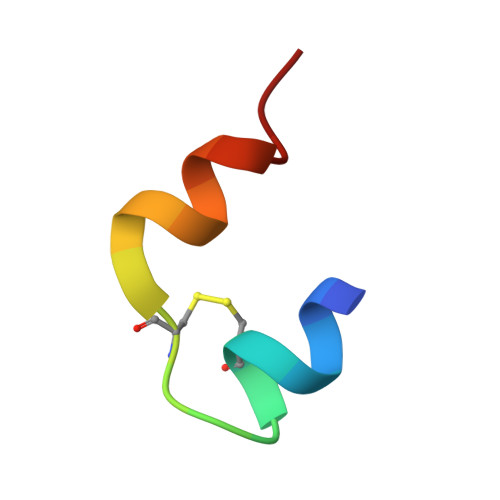
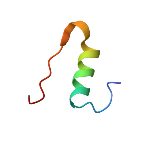
A review of the strategies for obtaining high-quality crystals utilizing nanotechnologies and microgravity

Pechkova, E.Bragazzi, N.Bozdaganyan, M.Belmonte, L.Nicolini, C.

(2014) Crit Rev Eukaryot Gene Expr 24: 325-339

  • DOI: https://doi.org/10.1615/critreveukaryotgeneexpr.2014008275
  • Primary Citation Related Structures: 
    4I5Z

  • PubMed Abstract: 

    Crystallization is a highly demanding and time-consuming task that causes a real bottle-neck in basic research. Great effort has been made to understand the factors and parameters that influence this process and to finely tune them to facilitate crystal growth. Different crystallization techniques have been proposed over the past decades, such as the classical vapor hanging drop method, its variant the sitting drop method, dialysis, cryo-temperature, gel, batch, and the innovative microgravity (space) techniques like free interface diffusion (FID) and counter-ion diffusion (CID). Here, we present a review of the strategies utilizing Langmuir-Blodgett (LB)-based nanotechnologies, and microgravity techniques for obtaining optimal high-quality crystals, as proven by molecular dynamics (MD) and bioinformatics approaches, namely using a clustering algorithm and protein alignment.


  • Organizational Affiliation
    • Nanoworld Institute Fondazione ELBA Nicolini, Pradalunga, Bergamo 24100, Italy ; Biophysics and Nanobiotechnology Laboratories, Department of Experimental Medicine, University of Genova, Genoa 16121-16167, Italy; European Synchrotron Radiation Facility.

Macromolecule Content 

  • Total Structure Weight: 5.74 kDa 
  • Atom Count: 454 
  • Modeled Residue Count: 51 
  • Deposited Residue Count: 51 
  • Unique protein chains: 2

Macromolecules

Find similar proteins by:  Sequence   |   3D Structure  
Entity ID: 1
MoleculeChains  Sequence LengthOrganismDetailsImage
Insulin21Bos taurusMutation(s): 0 
Gene Names: INS
UniProt
Find proteins for P01317 (Bos taurus)
Explore P01317 
Go to UniProtKB:  P01317
Entity Groups
Sequence Clusters30% Identity50% Identity70% Identity90% Identity95% Identity100% Identity
UniProt GroupP01317
Sequence Annotations
Expand
Reference Sequence
Find similar proteins by:|  3D Structure
Entity ID: 2
MoleculeChains  Sequence LengthOrganismDetailsImage
Insulin30Bos taurusMutation(s): 0 
Gene Names: INS
UniProt
Find proteins for P01317 (Bos taurus)
Explore P01317 
Go to UniProtKB:  P01317
Entity Groups
Sequence Clusters30% Identity50% Identity70% Identity90% Identity95% Identity100% Identity
UniProt GroupP01317
Sequence Annotations
Expand
Reference Sequence

Experimental Data & Validation

Experimental Data

  • Method: X-RAY DIFFRACTION
  • Resolution: 1.80 Å
  • R-Value Free:  0.218 (Depositor), 0.213 (DCC) 
  • R-Value Work:  0.179 (Depositor), 0.179 (DCC) 
  • R-Value Observed: 0.181 (Depositor) 
Space Group: I 21 3
Unit Cell:
Length ( Å )Angle ( ˚ )
a = 78.22α = 90
b = 78.22β = 90
c = 78.22γ = 90
Software Package:
Software NamePurpose
MOSFLMdata reduction
SCALAdata scaling
MOLREPphasing
REFMACrefinement
PDB_EXTRACTdata extraction

Structure Validation

View Full Validation Report


Entry History 

Deposition Data

Revision History  (Full details and data files)

  • Version 1.0: 2013-11-20
    Type: Initial release
  • Version 1.1: 2015-12-09
    Changes: Database references
  • Version 1.2: 2015-12-16
    Changes: Other
  • Version 1.3: 2024-10-30
    Changes: Data collection, Database references, Structure summary