4I47

Crystal structure of the Ribosome inactivating protein complexed with methylated guanine


Experimental Data Snapshot

  • Method: X-RAY DIFFRACTION
  • Resolution: 2.65 Å
  • R-Value Free: 0.199 
  • R-Value Work: 0.165 
  • R-Value Observed: 0.167 

wwPDB Validation   3D Report Full Report


Ligand Structure Quality Assessment 


This is version 2.1 of the entry. See complete history


Literature

First structural evidence of sequestration of mRNA cap structures by type 1 ribosome inactivating protein from Momordica balsamina.

Kushwaha, G.S.Yamini, S.Kumar, M.Sinha, M.Kaur, P.Sharma, S.Singh, T.P.

(2013) Proteins 81: 896-905

  • DOI: https://doi.org/10.1002/prot.24248
  • Primary Citation of Related Structures:  
    4EMF, 4EMR, 4I47

  • PubMed Abstract: 

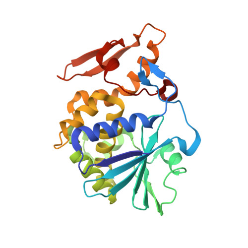
    This is the first structural evidence of recognition of mRNA cap structures by a ribosome inactivating protein. It is well known that a unique cap structure is formed at the 5' end of mRNA for carrying out various processes including mRNA maturation, translation initiation, and RNA turnover. The binding studies and crystal structure determinations of type 1 ribosome inactivating protein (RIP-1) from Momordica balsamina (MbRIP-1) were carried out with mRNA cap structures including (i) N7-methyl guanine (m7G), (ii) N7-methyl guanosine diphosphate (m7GDP), and (iii) N7-methyl guanosine triphosphate (m7GTP). These compounds showed affinities to MbRIP-1 at nanomolar concentrations. The structure determinations of the complexes of MbRIP-1 with m7G, m7GDP, and m7GTP at 2.65, 1.77, and 1.75 Å resolutions revealed that all the three compounds bound to MbRIP-1 in the substrate binding site at the positions which are slightly shifted towards Glu85 as compared to those of rRNA substrates. In this position, Glu85 forms several hydrogen bonds with guanine moiety while N-7 methyl group forms van der Waals contacts. However, the guanine rings are poorly stacked in these complexes. Thus, the mode of binding by MbRIP-1 to mRNA cap structures is different which results in the inhibition of depurination. Since some viruses are known to exploit the capping property of the host, this action of MbRIP-1 may have implications for the antiviral activity of this protein in vivo. The understanding of the mode of binding of MbRIP-1 to cap structures may also assist in the design of anti-viral agents.


  • Organizational Affiliation

    Department of Biophysics, All India Institute of Medical Sciences, New Delhi, India.


Macromolecules
Find similar proteins by:  (by identity cutoff)  |  3D Structure
Entity ID: 1
MoleculeChains Sequence LengthOrganismDetailsImage
rRNA N-glycosidase246Momordica balsaminaMutation(s): 0 
EC: 3.2.2.22
UniProt
Find proteins for D9J2T9 (Momordica balsamina)
Explore D9J2T9 
Go to UniProtKB:  D9J2T9
Entity Groups  
Sequence Clusters30% Identity50% Identity70% Identity90% Identity95% Identity100% Identity
UniProt GroupD9J2T9
Sequence Annotations
Expand
  • Reference Sequence
Oligosaccharides

Help

Entity ID: 2
MoleculeChains Length2D Diagram Glycosylation3D Interactions
2-acetamido-2-deoxy-beta-D-glucopyranose-(1-4)-2-acetamido-2-deoxy-beta-D-glucopyranose
B
2N-Glycosylation
Glycosylation Resources
GlyTouCan:  G42666HT
GlyCosmos:  G42666HT
GlyGen:  G42666HT
Small Molecules
Ligands 1 Unique
IDChains Name / Formula / InChI Key2D Diagram3D Interactions
MY6
Query on MY6

Download Ideal Coordinates CCD File 
C [auth A]2-amino-7-methyl-1,7-dihydro-6H-purin-6-one
C6 H7 N5 O
FZWGECJQACGGTI-UHFFFAOYSA-N
Binding Affinity Annotations 
IDSourceBinding Affinity
MY6 PDBBind:  4I47 Kd: 120 (nM) from 1 assay(s)
Experimental Data & Validation

Experimental Data

  • Method: X-RAY DIFFRACTION
  • Resolution: 2.65 Å
  • R-Value Free: 0.199 
  • R-Value Work: 0.165 
  • R-Value Observed: 0.167 
  • Space Group: H 3
Unit Cell:
Length ( Å )Angle ( ˚ )
a = 131.428α = 90
b = 131.428β = 90
c = 39.808γ = 120
Software Package:
Software NamePurpose
AMoREphasing
REFMACrefinement
AUTOMARdata reduction
SCALEPACKdata scaling

Structure Validation

View Full Validation Report


Ligand Structure Quality Assessment 


Entry History 

Deposition Data

Revision History  (Full details and data files)

  • Version 1.0: 2012-12-12
    Type: Initial release
  • Version 1.1: 2013-05-15
    Changes: Database references
  • Version 2.0: 2020-07-29
    Type: Remediation
    Reason: Carbohydrate remediation
    Changes: Advisory, Atomic model, Data collection, Derived calculations, Structure summary
  • Version 2.1: 2023-11-08
    Changes: Data collection, Database references, Refinement description, Structure summary