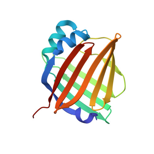
4I3B

Crystal structure of fluorescent protein UnaG wild type


Experimental Data Snapshot

  • Method: X-RAY DIFFRACTION
  • Resolution: 1.20 Å
  • R-Value Free: 0.159 
  • R-Value Work: 0.129 
  • R-Value Observed: 0.131 

wwPDB Validation   3D Report Full Report


Ligand Structure Quality Assessment 


This is version 1.3 of the entry. See complete history


Literature

A bilirubin-inducible fluorescent protein from eel muscle

Kumagai, A.Ando, R.Miyatake, H.Greimel, P.Kobayashi, T.Hirabayashi, Y.Shimogori, T.Miyawaki, A.

(2013) Cell 153: 1602-1611

  • DOI: https://doi.org/10.1016/j.cell.2013.05.038
  • Primary Citation of Related Structures:  
    4I3B, 4I3C, 4I3D

  • PubMed Abstract: 

    The fluorescent protein toolbox has revolutionized experimental biology. Despite this advance, no fluorescent proteins have been identified from vertebrates, nor has chromogenic ligand-inducible activation or clinical utility been demonstrated. Here, we report the cloning and characterization of UnaG, a fluorescent protein from Japanese eel. UnaG belongs to the fatty-acid-binding protein (FABP) family, and expression in eel is restricted to small-diameter muscle fibers. On heterologous expression in cell lines or mouse brain, UnaG produces oxygen-independent green fluorescence. Remarkably, UnaG fluorescence is triggered by an endogenous ligand, bilirubin, a membrane-permeable heme metabolite and clinical health biomarker. The holoUnaG structure at 1.2 Å revealed a biplanar coordination of bilirubin by reversible π-conjugation, and we used this high-affinity and high-specificity interaction to establish a fluorescence-based human bilirubin assay with promising clinical utility. UnaG will be the prototype for a versatile class of ligand-activated fluorescent proteins, with applications in research, medicine, and bioengineering.


  • Organizational Affiliation

    Cell Function Dynamics, Brain Science Institute, RIKEN, 2-1 Hirosawa, Wako-city, Saitama 351-0198, Japan.


Macromolecules
Find similar proteins by:  (by identity cutoff)  |  3D Structure
Entity ID: 1
MoleculeChains Sequence LengthOrganismDetailsImage
Bilirubin-inducible fluorescent protein UnaG
A, B, C, D, E
A, B, C, D, E, F
139Anguilla japonicaMutation(s): 0 
UniProt
Find proteins for P0DM59 (Anguilla japonica)
Explore P0DM59 
Go to UniProtKB:  P0DM59
Entity Groups  
Sequence Clusters30% Identity50% Identity70% Identity90% Identity95% Identity100% Identity
UniProt GroupP0DM59
Sequence Annotations
Expand
  • Reference Sequence
Small Molecules
Ligands 2 Unique
IDChains Name / Formula / InChI Key2D Diagram3D Interactions
BLR
Query on BLR

Download Ideal Coordinates CCD File 
G [auth A]
I [auth B]
M [auth C]
N [auth D]
P [auth E]
G [auth A],
I [auth B],
M [auth C],
N [auth D],
P [auth E],
R [auth F]
3-[5-[(Z)-(4-ethenyl-3-methyl-5-oxidanylidene-pyrrol-2-ylidene)methyl]-2-[[5-[(Z)-(3-ethenyl-4-methyl-5-oxidanylidene-pyrrol-2-ylidene)methyl]-3-(3-hydroxy-3-oxopropyl)-4-methyl-1H-pyrrol-2-yl]methyl]-4-methyl-1H-pyrrol-3-yl]propanoic acid
C33 H36 N4 O6
BPYKTIZUTYGOLE-IFADSCNNSA-N
PEG
Query on PEG

Download Ideal Coordinates CCD File 
H [auth A]
J [auth B]
K [auth B]
L [auth B]
O [auth D]
H [auth A],
J [auth B],
K [auth B],
L [auth B],
O [auth D],
Q [auth E],
S [auth F],
T [auth F],
U [auth F]
DI(HYDROXYETHYL)ETHER
C4 H10 O3
MTHSVFCYNBDYFN-UHFFFAOYSA-N
Experimental Data & Validation

Experimental Data

  • Method: X-RAY DIFFRACTION
  • Resolution: 1.20 Å
  • R-Value Free: 0.159 
  • R-Value Work: 0.129 
  • R-Value Observed: 0.131 
  • Space Group: P 1 21 1
Unit Cell:
Length ( Å )Angle ( ˚ )
a = 44.394α = 90
b = 73.954β = 92.45
c = 124.364γ = 90
Software Package:
Software NamePurpose
HKL-2000data collection
PHENIXmodel building
PHENIXrefinement
HKL-2000data reduction
HKL-2000data scaling
PHENIXphasing

Structure Validation

View Full Validation Report


Ligand Structure Quality Assessment 


Entry History 

Deposition Data

Revision History  (Full details and data files)

  • Version 1.0: 2013-06-19
    Type: Initial release
  • Version 1.1: 2013-07-31
    Changes: Database references
  • Version 1.2: 2015-02-04
    Changes: Database references, Non-polymer description
  • Version 1.3: 2023-11-08
    Changes: Data collection, Database references, Derived calculations, Refinement description