4HZ6 | pdb_00004hz6

crystal structure of BglB


Experimental Data Snapshot

  • Method: X-RAY DIFFRACTION
  • Resolution: 1.40 Å
  • R-Value Free: 
    0.179 (Depositor), 0.174 (DCC) 
  • R-Value Work: 
    0.138 (Depositor), 0.134 (DCC) 
  • R-Value Observed: 
    0.142 (Depositor) 

Starting Model: experimental
View more details

wwPDB Validation 3D Report Full Report

Validation slider image for 4HZ6

This is version 1.1 of the entry. See complete history

Literature

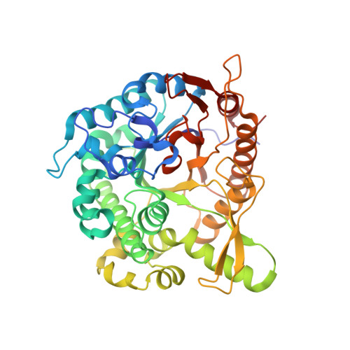
Structural insights into the substrate recognition properties of beta-glucosidase.

Nam, K.H.Sung, M.W.Hwang, K.Y.

(2010) Biochem Biophys Res Commun 391: 1131-1135

  • DOI: https://doi.org/10.1016/j.bbrc.2009.12.038
  • Primary Citation Related Structures: 
    4HZ6, 4HZ7, 4HZ8

  • PubMed Abstract: 

    Beta-glucosidase enzymes (EC 3.2.1-3.2.3) hydrolyze sugars and are implicated in a wide spectrum of biological processes. Recently, we reported that beta-glucosidase has varied kinetic parameters for the natural and synthetic substrates [K.H Nam, S.J. Kim, M.Y. Kim, J.H. Kim, T.S. Yeo, C.M. Lee, H.K Jun, K.Y. Hwang. Crystal structure of engineered beta-glucosidase from a soil metagenome, Proteins 73 (2008) 788-793]. However, an understanding of the kinetic values of beta-glucosidase has not yet enabled the elucidation of its molecular function. Here, we report the X-ray crystal structure of beta-glucosidase with a glucose and cellobiose fragment from uncultured soil metagenome. From the various crystals, we obtained the pre-reaction (native), intermediate (disaccharide cleavage) and post-reaction (glucose binding) states of the active site pocket. These structures provide snapshots of the catalytic processing of beta-glucosidase. In addition, the intermediate state of the crystal structure provides insight into the substrate specificity of beta-glucosidase. These structural studies will facilitate elucidation of the architectural mechanism responsible for the substrate recognition of beta-glucosidase.


  • Organizational Affiliation
    • Division of Biotechnology, College of Life Sciences & Biotechnology, Korea University, Seoul 136-701, South Korea.

Macromolecule Content 

  • Total Structure Weight: 50.39 kDa 
  • Atom Count: 3,980 
  • Modeled Residue Count: 441 
  • Deposited Residue Count: 444 
  • Unique protein chains: 1

Macromolecules

Find similar proteins by:|  3D Structure
Entity ID: 1
MoleculeChains  Sequence LengthOrganismDetailsImage
Beta-glucosidase444uncultured bacteriumMutation(s): 8 
Gene Names: bglA
UniProt
Find proteins for A0ACD6B8B8 (uncultured bacterium)
Explore A0ACD6B8B8 
Go to UniProtKB:  A0ACD6B8B8
Entity Groups
Sequence Clusters30% Identity50% Identity70% Identity90% Identity95% Identity100% Identity
UniProt GroupA0ACD6B8B8
Sequence Annotations
Expand
Reference Sequence

Small Molecules

Ligands 1 Unique
IDChains Name / Formula / InChI Key2D Diagram3D Interactions
GOL

Query on GOL


Download:Ideal Coordinates CCD File
B [auth A]GLYCEROL
C3 H8 O3
PEDCQBHIVMGVHV-UHFFFAOYSA-N

Experimental Data & Validation

Experimental Data

  • Method: X-RAY DIFFRACTION
  • Resolution: 1.40 Å
  • R-Value Free:  0.179 (Depositor), 0.174 (DCC) 
  • R-Value Work:  0.138 (Depositor), 0.134 (DCC) 
  • R-Value Observed: 0.142 (Depositor) 
Space Group: P 21 21 21
Unit Cell:
Length ( Å )Angle ( ˚ )
a = 70.697α = 90
b = 71.143β = 90
c = 87.373γ = 90
Software Package:
Software NamePurpose
REFMACrefinement
PDB_EXTRACTdata extraction
HKL-2000data collection
HKL-2000data reduction
HKL-2000data scaling
CNSphasing

Structure Validation

View Full Validation Report


Entry History 

Deposition Data

Revision History  (Full details and data files)

  • Version 1.0: 2012-12-19
    Type: Initial release
  • Version 1.1: 2023-11-08
    Changes: Data collection, Database references, Derived calculations, Refinement description