4HYH | pdb_00004hyh

X-RAY Crystal structure of compound 39 bound to human chk1 kinase domain


Experimental Data Snapshot

  • Method: X-RAY DIFFRACTION
  • Resolution: 1.70 Å
  • R-Value Free: 
    0.226 (Depositor), 0.265 (DCC) 
  • R-Value Work: 
    0.203 (Depositor) 
  • R-Value Observed: 
    0.204 (Depositor) 

wwPDB Validation 3D Report Full Report

Validation slider image for 4HYH

Ligand Structure Quality Assessment 


This is version 1.2 of the entry. See complete history

Literature

Structure-based design and optimization of 2-aminothiazole-4-carboxamide as a new class of CHK1 inhibitors.

Huang, X.Cheng, C.C.Fischmann, T.O.Duca, J.S.Richards, M.Tadikonda, P.K.Reddy, P.A.Zhao, L.Arshad Siddiqui, M.Parry, D.Davis, N.Seghezzi, W.Wiswell, D.Shipps, G.W.

(2013) Bioorg Med Chem Lett 23: 2590-2594

  • DOI: https://doi.org/10.1016/j.bmcl.2013.02.108
  • Primary Citation Related Structures: 
    4HYH, 4HYI

  • PubMed Abstract: 

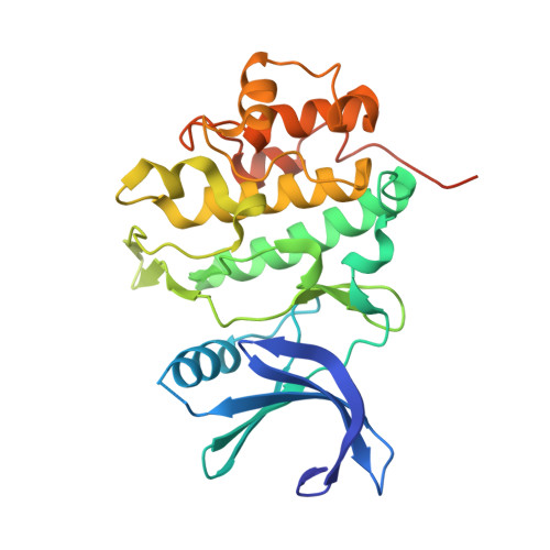
    Drug design efforts in the emerging 2-aminothiazole-4-carboxamide class of CHK1 inhibitors have uncovered specific combinations of key substructures within the molecule; resulting in significant improvements in cell-based activity while retaining a greater than one hundred-fold selectivity against CDK2. The X-ray crystal structure of a complex between compound 39 and the CHK1 protein detailing a 'U-shaped' topology and key interactions with the protein surface at the ATP site is also reported.


  • Organizational Affiliation
    • Merck Research Laboratories, 320 Bent Street, Cambridge, MA 02141, USA. xiaohua.huang@merck.com

Macromolecule Content 

  • Total Structure Weight: 33.49 kDa 
  • Atom Count: 2,297 
  • Modeled Residue Count: 263 
  • Deposited Residue Count: 289 
  • Unique protein chains: 1

Macromolecules

Find similar proteins by:|  3D Structure
Entity ID: 1
MoleculeChains  Sequence LengthOrganismDetailsImage
Serine/threonine-protein kinase Chk1289Homo sapiensMutation(s): 0 
Gene Names: CHEK1CHK1
EC: 2.7.11.1
UniProt & NIH Common Fund Data Resources
Find proteins for O14757 (Homo sapiens)
Explore O14757 
Go to UniProtKB:  O14757
PHAROS:  O14757
GTEx:  ENSG00000149554 
Entity Groups
Sequence Clusters30% Identity50% Identity70% Identity90% Identity95% Identity100% Identity
UniProt GroupO14757
Sequence Annotations
Expand
Reference Sequence

Small Molecules

Ligands 1 Unique
IDChains Name / Formula / InChI Key2D Diagram3D Interactions
1AM

Query on 1AM


Download:Ideal Coordinates CCD File
B [auth A]2-(6-methoxy-1-oxo-1,3-dihydro-2H-isoindol-2-yl)-N-[4-(piperazin-1-yl)pyridin-3-yl]-1,3-thiazole-4-carboxamide
C22 H22 N6 O3 S
KLJVDMAOKMSBQX-UHFFFAOYSA-N
Binding Affinity Annotations 
IDSourceBinding Affinity
1AM BindingDB:  4HYH IC50: min: 1, max: 3.7 (nM) from 2 assay(s)
EC50: 30 (nM) from 1 assay(s)

Experimental Data & Validation

Experimental Data

  • Method: X-RAY DIFFRACTION
  • Resolution: 1.70 Å
  • R-Value Free:  0.226 (Depositor), 0.265 (DCC) 
  • R-Value Work:  0.203 (Depositor) 
  • R-Value Observed: 0.204 (Depositor) 
Space Group: P 1 21 1
Unit Cell:
Length ( Å )Angle ( ˚ )
a = 44.97α = 90
b = 65.96β = 102.39
c = 54.42γ = 90
Software Package:
Software NamePurpose
CrystalCleardata collection
BUSTERrefinement
DENZOdata reduction
SCALEPACKdata scaling
BUSTERphasing

Structure Validation

View Full Validation Report


Ligand Structure Quality Assessment 


Entry History 

Deposition Data

Revision History  (Full details and data files)

  • Version 1.0: 2013-03-06
    Type: Initial release
  • Version 1.1: 2013-05-01
    Changes: Database references
  • Version 1.2: 2024-02-28
    Changes: Data collection, Database references, Derived calculations