4HSU | pdb_00004hsu

Crystal structure of LSD2-NPAC with H3(1-26)in space group P21


Experimental Data Snapshot

  • Method: X-RAY DIFFRACTION
  • Resolution: 1.99 Å
  • R-Value Free: 
    0.232 (Depositor), 0.233 (DCC) 
  • R-Value Work: 
    0.199 (Depositor), 0.200 (DCC) 
  • R-Value Observed: 
    0.201 (Depositor) 

Starting Model: experimental
View more details

wwPDB Validation 3D Report Full Report

Validation slider image for 4HSU

Ligand Structure Quality Assessment 


This is version 1.2 of the entry. See complete history

Literature

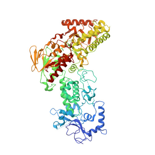
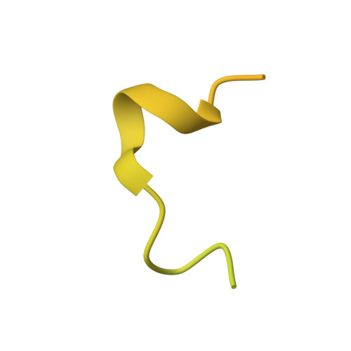
Structural insight into substrate recognition by histone demethylase LSD2/KDM1b.

Chen, F.Yang, H.Dong, Z.Fang, J.Wang, P.Zhu, T.Gong, W.Fang, R.Shi, Y.G.Li, Z.Xu, Y.

(2013) Cell Res 23: 306-309

Macromolecule Content 

  • Total Structure Weight: 104.74 kDa 
  • Atom Count: 6,505 
  • Modeled Residue Count: 775 
  • Deposited Residue Count: 930 
  • Unique protein chains: 3

Macromolecules

Find similar proteins by:|  3D Structure
Entity ID: 1
MoleculeChains  Sequence LengthOrganismDetailsImage
Lysine-specific histone demethylase 1B776Homo sapiensMutation(s): 0 
Gene Names: LSD2
EC: 1 (PDB Primary Data), 1.14.99.66 (UniProt)
UniProt & NIH Common Fund Data Resources
Find proteins for Q8NB78 (Homo sapiens)
Explore Q8NB78 
Go to UniProtKB:  Q8NB78
PHAROS:  Q8NB78
GTEx:  ENSG00000165097 
Entity Groups
Sequence Clusters30% Identity50% Identity70% Identity90% Identity95% Identity100% Identity
UniProt GroupQ8NB78
Sequence Annotations
Expand
Reference Sequence
Find similar proteins by:|  3D Structure
Entity ID: 2
MoleculeChains  Sequence LengthOrganismDetailsImage
Putative oxidoreductase GLYR1124Homo sapiensMutation(s): 0 
Gene Names: GLYR1
EC: 1
UniProt & NIH Common Fund Data Resources
Find proteins for Q49A26 (Homo sapiens)
Explore Q49A26 
Go to UniProtKB:  Q49A26
PHAROS:  Q49A26
GTEx:  ENSG00000140632 
Entity Groups
Sequence Clusters30% Identity50% Identity70% Identity90% Identity95% Identity100% Identity
UniProt GroupQ49A26
Sequence Annotations
Expand
Reference Sequence
Find similar proteins by:|  3D Structure
Entity ID: 3
MoleculeChains  Sequence LengthOrganismDetailsImage
Histone H330Xenopus laevisMutation(s): 1 
UniProt
Find proteins for Q92133 (Xenopus laevis)
Explore Q92133 
Go to UniProtKB:  Q92133
Entity Groups
Sequence Clusters30% Identity50% Identity70% Identity90% Identity95% Identity100% Identity
UniProt GroupQ92133
Sequence Annotations
Expand
Reference Sequence

Experimental Data & Validation

Experimental Data

  • Method: X-RAY DIFFRACTION
  • Resolution: 1.99 Å
  • R-Value Free:  0.232 (Depositor), 0.233 (DCC) 
  • R-Value Work:  0.199 (Depositor), 0.200 (DCC) 
  • R-Value Observed: 0.201 (Depositor) 
Space Group: P 1 21 1
Unit Cell:
Length ( Å )Angle ( ˚ )
a = 62.754α = 90
b = 89.481β = 102.89
c = 88.349γ = 90
Software Package:
Software NamePurpose
HKL-2000data collection
PHASERphasing
PHENIXrefinement
HKL-2000data reduction
HKL-2000data scaling

Structure Validation

View Full Validation Report


Ligand Structure Quality Assessment 


Entry History 

Deposition Data

Revision History  (Full details and data files)

  • Version 1.0: 2013-02-13
    Type: Initial release
  • Version 1.1: 2013-07-10
    Changes: Database references
  • Version 1.2: 2023-11-08
    Changes: Data collection, Database references, Derived calculations, Refinement description