Structural studies of Streptococcus pyogenes streptolysin O provide insights into the early steps of membrane penetration.
Feil, S.C., Ascher, D.B., Kuiper, M.J., Tweten, R.K., Parker, M.W.(2014) J Mol Biology 426: 785-792
- PubMed: 24316049 Search on PubMedSearch on PubMed Central
- DOI: https://doi.org/10.1016/j.jmb.2013.11.020
- Primary Citation Related Structures:
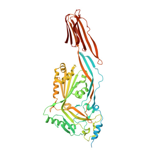
4HSC - PubMed Abstract:
Cholesterol-dependent cytolysins (CDCs) are a large family of bacterial toxins that exhibit a dependence on the presence of membrane cholesterol in forming large pores in cell membranes. Significant changes in the three-dimensional structure of these toxins are necessary to convert the soluble monomeric protein into a membrane pore. We have determined the crystal structure of the archetypical member of the CDC family, streptolysin O (SLO), a virulence factor from Streptococcus pyogenes. The overall fold is similar to previously reported CDC structures, although the C-terminal domain is in a different orientation with respect to the rest of the molecule. Surprisingly, a signature stretch of CDC sequence called the undecapeptide motif, a key region involved in membrane recognition, adopts a very different structure in SLO to that of the well-characterized CDC perfringolysin O (PFO), although the sequences in this region are identical. An analysis reveals that, in PFO, there are complementary interactions between the motif and the rest of domain 4 that are lost in SLO. Molecular dynamics simulations suggest that the loss of a salt bridge in SLO and a cation-pi interaction are determining factors in the extended conformation of the motif, which in turn appears to result in a greater flexibility of the neighboring L1 loop that houses a cholesterol-sensing motif. These differences may explain the differing abilities of SLO and PFO to efficiently penetrate target cell membranes in the first step of toxin insertion into the membrane.
- ACRF Rational Drug Discovery Centre, Biota Structural Biology Laboratory, St. Vincent's Institute of Medical Research, Fitzroy, Victoria 3065, Australia.
Organizational Affiliation: