4HQE | pdb_00004hqe

The crystal structure of QsrR-DNA complex


Experimental Data Snapshot

  • Method: X-RAY DIFFRACTION
  • Resolution: 2.30 Å
  • R-Value Free: 
    0.260 (Depositor), 0.250 (DCC) 
  • R-Value Work: 
    0.205 (Depositor), 0.197 (DCC) 
  • R-Value Observed: 
    0.208 (Depositor) 

wwPDB Validation 3D Report Full Report

Validation slider image for 4HQE

This is version 1.2 of the entry. See complete history

Literature

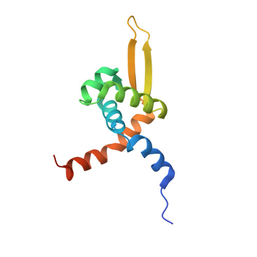
Molecular mechanism of quinone signaling mediated through S-quinonization of a YodB family repressor QsrR.

Ji, Q.Zhang, L.Jones, M.B.Sun, F.Deng, X.Liang, H.Cho, H.Brugarolas, P.Gao, Y.N.Peterson, S.N.Lan, L.Bae, T.He, C.

(2013) Proc Natl Acad Sci U S A 110: 5010-5015

  • DOI: https://doi.org/10.1073/pnas.1219446110
  • Primary Citation Related Structures: 
    4HQE, 4HQM

  • PubMed Abstract: 

    Quinone molecules are intracellular electron-transport carriers, as well as critical intra- and extracellular signals. However, transcriptional regulation of quinone signaling and its molecular basis are poorly understood. Here, we identify a thiol-stress-sensing regulator YodB family transcriptional regulator as a central component of quinone stress response of Staphylococcus aureus, which we have termed the quinone-sensing and response repressor (QsrR). We also identify and confirm an unprecedented quinone-sensing mechanism based on the S-quinonization of the essential residue Cys-5. Structural characterizations of the QsrR-DNA and QsrR-menadione complexes further reveal that the covalent association of menadione directly leads to the release of QsrR from operator DNA following a 10° rigid-body rotation as well as a 9-Å elongation between the dimeric subunits. The molecular level characterization of this quinone-sensing transcriptional regulator provides critical insights into quinone-mediated gene regulation in human pathogens.


  • Organizational Affiliation
    • Department of Chemistry, The University of Chicago, Chicago, IL 60637, USA.

Macromolecule Content 

  • Total Structure Weight: 36.62 kDa 
  • Atom Count: 2,498 
  • Modeled Residue Count: 247 
  • Deposited Residue Count: 264 
  • Unique protein chains: 1
  • Unique nucleic acid chains: 2

Macromolecules


Find similar proteins by:|  3D Structure
Entity ID: 1
MoleculeChains  Sequence LengthOrganismDetailsImage
Transcriptional regulator QsrR
A, B
115Staphylococcus aureusMutation(s): 0 
Gene Names: SAV2123
UniProt
Find proteins for A0A0H3JWI0 (Staphylococcus aureus (strain Mu50 / ATCC 700699))
Explore A0A0H3JWI0 
Go to UniProtKB:  A0A0H3JWI0
Entity Groups
Sequence Clusters30% Identity50% Identity70% Identity90% Identity95% Identity100% Identity
UniProt GroupA0A0H3JWI0
Sequence Annotations
Expand
Reference Sequence
Find similar nucleic acids by:  Sequence
Entity ID: 2
MoleculeChains LengthOrganismImage
DNA (5'-D(*GP*GP*TP*AP*TP*AP*AP*TP*AP*AP*TP*TP*AP*TP*AP*CP*T)-3')17synthetic construct
Sequence Annotations
Expand
Reference Sequence
Find similar nucleic acids by:  Sequence
Entity ID: 3
MoleculeChains LengthOrganismImage
DNA (5'-D(*AP*GP*TP*AP*TP*AP*AP*TP*TP*AP*TP*TP*AP*TP*AP*CP*C)-3')17synthetic construct
Sequence Annotations
Expand
Reference Sequence

Experimental Data & Validation

Experimental Data

  • Method: X-RAY DIFFRACTION
  • Resolution: 2.30 Å
  • R-Value Free:  0.260 (Depositor), 0.250 (DCC) 
  • R-Value Work:  0.205 (Depositor), 0.197 (DCC) 
  • R-Value Observed: 0.208 (Depositor) 
Space Group: P 41
Unit Cell:
Length ( Å )Angle ( ˚ )
a = 48.189α = 90
b = 48.189β = 90
c = 156.807γ = 90
Software Package:
Software NamePurpose
HKL-2000data collection
PHASESphasing
PHENIXrefinement
HKL-2000data reduction
HKL-2000data scaling

Structure Validation

View Full Validation Report


Entry History 

Deposition Data

Revision History  (Full details and data files)

  • Version 1.0: 2013-03-06
    Type: Initial release
  • Version 1.1: 2013-05-22
    Changes: Database references
  • Version 1.2: 2024-02-28
    Changes: Data collection, Database references