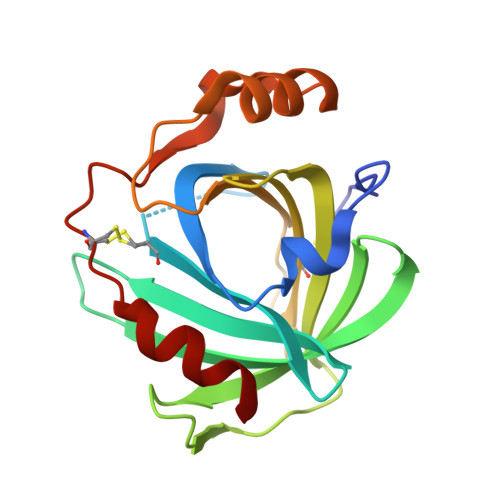
Complexes of ferriheme nitrophorin 4 with low-molecular weight thiol(ate)s occurring in blood plasma
He, C., Nishikawa, K., Erdem, O.F., Reijerse, E., Ogata, H., Lubitz, W., Knipp, M.(2013) J Inorg Biochem 122: 38-48
- PubMed: 23474537
- DOI: https://doi.org/10.1016/j.jinorgbio.2013.01.012
- Primary Citation of Related Structures:
4HPA, 4HPB, 4HPC, 4HPD - PubMed Abstract:
Nitrophorins are proteins occurring in the saliva of the blood-sucking insect Rhodnius prolixus to carry NO as a vasodilator and blood-coagulation inhibitor into the victim's tissue. It was suggested that the rate of NO release can be enhanced by the blood-plasma component L-cysteine [J.M.C.Ribeiro, Insect Biochem. Mol. Biol. 26 (1996) 899-905]. However, the mechanism of the reaction is not clear. In the attempt to exploit the reaction in detail, complexes of nitrophorin 4 (NP4) with the thiols 2-mercaptoethanol, L-cysteine, and L-homocysteine and with HS(-) were formed and characterized under anaerobic conditions using absorption spectroscopy, X-ray crystallography, and EPR spectroscopy. In contrast to met-myoglobin, which is reduced by L-cysteine, all four compounds form low-spin Fe(III) complexes with NP4. The weak equilibration constants (167-5200 M(-1)) neither support significant complexation nor the simple displacement of NO in vivo. Both amino acid based thiols form additional H-bonds with side chains of the heme pocket entry. Glutathione and L-methionine did not form a complex, indicating the specificity of the complexes with L-cysteine and L-homocysteine. Continuous wave EPR spectroscopy reveals the simultaneous existence of three low-spin systems in each case that are attributed to various protonation and/or conformational stages in the heme pocket. Electron nuclear double resonance (ENDOR) spectroscopy demonstrates that the thiol sulfurs are, at least in part, protonated. Overall, the results not only demonstrate the good accessibility of the NP4 heme center by biologically relevant thiols, but also represent the first structural characterization of a ferriheme protein in complex with L-cysteine L-homocysteine.
- Max-Planck-Institut für Chemische Energiekonversion, Stiftstrasse 34-36, D-45470 Mülheim an der Ruhr, Germany.
Organizational Affiliation:


