Structural and Biochemical Analyses of the Eukaryotic Heat Shock Locus V (HslV) from Trypanosoma brucei.
Sung, K.H., Lee, S.Y., Song, H.K.(2013) J Biol Chem 288: 23234-23243
- PubMed: 23818520 Search on PubMedSearch on PubMed Central
- DOI: https://doi.org/10.1074/jbc.M113.484832
- Primary Citation Related Structures:
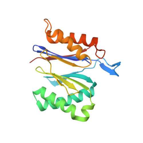
4HNZ, 4HO7 - PubMed Abstract:
In many bacteria, heat shock locus V (HslV) functions as a protease, which is activated by heat shock locus U (HslU). The primary sequence and structure of HslV are well conserved with those of the β-subunit of the 20 S proteasome core particle in eukaryotes. To date, the HslVU complex has only been characterized in the prokaryotic system. Recently, however, the coexistence of a 20 S proteasome with HslV protease in the same living organism has been reported. In Trypanosoma brucei, a protozoan parasite that causes human sleeping sickness in Africa, HslV is localized in the mitochondria, where it has a novel function in regulating mitochondrial DNA replication. Although the prokaryotic HslVU system has been studied extensively, little is known regarding its eukaryotic counterpart. Here, we report the biochemical characteristics of an HslVU complex from T. brucei. In contrast to the prokaryotic system, T. brucei possesses two potential HslU molecules, and we found that only one of them activates HslV. A key activating residue, Tyr(494), was identified in HslU2 by biochemical and mutational studies. Furthermore, to our knowledge, this study is the first to report the crystal structure of a eukaryotic HslV, determined at 2.4 Å resolution. Drawing on our comparison of the biochemical and structural data, we discuss herein the differences and similarities between eukaryotic and prokaryotic HslVs.
- Department of Life Sciences, Korea University, Anam-Dong, Seongbuk-Gu, Seoul 136-701, Korea.
Organizational Affiliation: