4HMQ

Crystal structure of streptococcus pneumoniae TIGR4 PiaA in complex with ferrichrome


Experimental Data Snapshot

  • Method: X-RAY DIFFRACTION
  • Resolution: 2.10 Å
  • R-Value Free: 0.248 
  • R-Value Work: 0.200 
  • R-Value Observed: 0.202 

wwPDB Validation   3D Report Full Report


Ligand Structure Quality Assessment 


This is version 1.1 of the entry. See complete history


Literature

Structures of Streptococcus pneumoniae PiaA and Its Complex with Ferrichrome Reveal Insights into the Substrate Binding and Release of High Affinity Iron Transporters

Cheng, W.Li, Q.Jiang, Y.-L.Zhou, C.-Z.Chen, Y.

(2013) PLoS One 8: e71451-e71451

  • DOI: https://doi.org/10.1371/journal.pone.0071451
  • Primary Citation of Related Structures:  
    4HMO, 4HMP, 4HMQ

  • PubMed Abstract: 

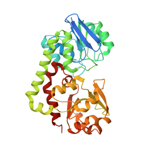
    Iron scarcity is one of the nutrition limitations that the Gram-positive infectious pathogens Streptococcus pneumoniae encounter in the human host. To guarantee sufficient iron supply, the ATP binding cassette (ABC) transporter Pia is employed to uptake iron chelated by hydroxamate siderophore, via the membrane-anchored substrate-binding protein PiaA. The high affinity towards ferrichrome enables PiaA to capture iron at a very low concentration in the host. We presented here the crystal structures of PiaA in both apo and ferrichrome-complexed forms at 2.7 and 2.1 Å resolution, respectively. Similar to other class III substrate binding proteins, PiaA is composed of an N-terminal and a C-terminal domain bridged by an α-helix. At the inter-domain cleft, a molecule of ferrichrome is stabilized by a number of highly conserved residues. Upon ferrichrome binding, two highly flexible segments at the entrance of the cleft undergo significant conformational changes, indicating their contribution to the binding and/or release of ferrichrome. Superposition to the structure of Escherichia coli ABC transporter BtuF enabled us to define two conserved residues: Glu119 and Glu262, which were proposed to form salt bridges with two arginines of the permease subunits. Further structure-based sequence alignment revealed that the ferrichrome binding pattern is highly conserved in a series of PiaA homologs encoded by both Gram-positive and negative bacteria, which were predicted to be sensitive to albomycin, a sideromycin antibiotic derived from ferrichrome.


  • Organizational Affiliation

    Hefei National Laboratory for Physical Sciences at the Microscale and School of Life Sciences, University of Science and Technology of China, Hefei, China.


Macromolecules
Find similar proteins by:  (by identity cutoff)  |  3D Structure
Entity ID: 1
MoleculeChains Sequence LengthOrganismDetailsImage
Iron-compound ABC transporter, iron compound-binding protein
A, B
328Streptococcus pneumoniae TIGR4Mutation(s): 0 
Gene Names: SP_1032
UniProt
Find proteins for A0A0H2UPT5 (Streptococcus pneumoniae serotype 4 (strain ATCC BAA-334 / TIGR4))
Explore A0A0H2UPT5 
Go to UniProtKB:  A0A0H2UPT5
Entity Groups  
Sequence Clusters30% Identity50% Identity70% Identity90% Identity95% Identity100% Identity
UniProt GroupA0A0H2UPT5
Sequence Annotations
Expand
  • Reference Sequence
Small Molecules
Ligands 5 Unique
IDChains Name / Formula / InChI Key2D Diagram3D Interactions
FCE
Query on FCE

Download Ideal Coordinates CCD File 
C [auth A],
S [auth B]
FERRICHROME
C27 H42 Fe N9 O12
GGUNGDGGXMHBMJ-OCIDDWSYSA-N
PGE
Query on PGE

Download Ideal Coordinates CCD File 
LA [auth B],
R [auth A]
TRIETHYLENE GLYCOL
C6 H14 O4
ZIBGPFATKBEMQZ-UHFFFAOYSA-N
CD
Query on CD

Download Ideal Coordinates CCD File 
AA [auth B]
BA [auth B]
CA [auth B]
D [auth A]
DA [auth B]
AA [auth B],
BA [auth B],
CA [auth B],
D [auth A],
DA [auth B],
E [auth A],
EA [auth B],
F [auth A],
FA [auth B],
G [auth A],
H [auth A],
I [auth A],
J [auth A],
K [auth A],
L [auth A],
M [auth A],
N [auth A],
T [auth B],
U [auth B],
V [auth B],
W [auth B],
X [auth B],
Y [auth B],
Z [auth B]
CADMIUM ION
Cd
WLZRMCYVCSSEQC-UHFFFAOYSA-N
EDO
Query on EDO

Download Ideal Coordinates CCD File 
IA [auth B],
JA [auth B],
KA [auth B],
P [auth A],
Q [auth A]
1,2-ETHANEDIOL
C2 H6 O2
LYCAIKOWRPUZTN-UHFFFAOYSA-N
CL
Query on CL

Download Ideal Coordinates CCD File 
GA [auth B],
HA [auth B],
O [auth A]
CHLORIDE ION
Cl
VEXZGXHMUGYJMC-UHFFFAOYSA-M
Binding Affinity Annotations 
IDSourceBinding Affinity
FCE PDBBind:  4HMQ Kd: 5.8 (nM) from 1 assay(s)
Experimental Data & Validation

Experimental Data

  • Method: X-RAY DIFFRACTION
  • Resolution: 2.10 Å
  • R-Value Free: 0.248 
  • R-Value Work: 0.200 
  • R-Value Observed: 0.202 
  • Space Group: P 21 21 21
Unit Cell:
Length ( Å )Angle ( ˚ )
a = 76.102α = 90
b = 84.272β = 90
c = 93.776γ = 90
Software Package:
Software NamePurpose
ADSCdata collection
AMoREphasing
REFMACrefinement
HKL-2000data reduction
SCALAdata scaling

Structure Validation

View Full Validation Report


Ligand Structure Quality Assessment 


Entry History 

Deposition Data

Revision History  (Full details and data files)

  • Version 1.0: 2013-09-04
    Type: Initial release
  • Version 1.1: 2024-03-20
    Changes: Data collection, Database references, Derived calculations