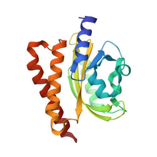
4HIA | pdb_00004hia

Crystal Structure of Rhodobacter Sphaeroides LOV protein


Experimental Data Snapshot

  • Method: X-RAY DIFFRACTION
  • Resolution: 1.95 Å
  • R-Value Free: 
    0.201 (Depositor), 0.200 (DCC) 
  • R-Value Work: 
    0.173 (Depositor), 0.180 (DCC) 
  • R-Value Observed: 
    0.175 (Depositor) 

wwPDB Validation   3D Report Full Report


Ligand Structure Quality Assessment 

Created with Raphaël 2.3.0Worse 01 BetterLigand structure goodness of fit to experimental dataBest fitted FMNClick on this verticalbar to view details

This is version 1.3 of the entry. See complete history


Literature

Light-induced subunit dissociation by a light-oxygen-voltage domain photoreceptor from Rhodobacter sphaeroides.

Conrad, K.S.Bilwes, A.M.Crane, B.R.

(2013) Biochemistry 52: 378-391

  • DOI: https://doi.org/10.1021/bi3015373
  • Primary Citation of Related Structures:  
    4HIA, 4HJ3, 4HJ4, 4HJ6, 4HNB

  • PubMed Abstract: 

    Light-oxygen-voltage (LOV) domains bind a flavin chromophore to serve as blue light sensors in a wide range of eukaryotic and prokaryotic proteins. LOV domains are associated with a variable effector domain or a separate protein signaling partner to execute a wide variety of functions that include regulation of kinases, generation of anti-sigma factor antagonists, and regulation of circadian clocks. Here we present the crystal structure, photocycle kinetics, association properties, and spectroscopic features of a full-length LOV domain protein from Rhodobacter sphaeroides (RsLOV). RsLOV exhibits N- and C-terminal helical extensions that form an unusual helical bundle at its dimer interface with some resemblance to the helical transducer of sensory rhodopsin II. The blue light-induced conformational changes of RsLOV revealed from a comparison of light- and dark-state crystal structures support a shared signaling mechanism of LOV domain proteins that originates with the light-induced formation of a flavin-cysteinyl photoadduct. Adduct formation disrupts hydrogen bonding in the active site and propagates structural changes through the LOV domain core to the N- and C-terminal extensions. Single-residue variants in the active site and dimer interface of RsLOV alter photoadduct lifetimes and induce structural changes that perturb the oligomeric state. Size exclusion chromatography, multiangle light scattering, small-angle X-ray scattering, and cross-linking studies indicate that RsLOV dimerizes in the dark but, upon light excitation, dissociates into monomers. This light-induced switch in oligomeric state may prove to be useful for engineering molecular associations in controlled cellular settings.


  • Organizational Affiliation

    Department of Chemistry and Chemical Biology, Cornell University, Ithaca, New York 14853, USA.


Macromolecules
Find similar proteins by:  (by identity cutoff)  |  3D Structure
Entity ID: 1
MoleculeChains Sequence LengthOrganismDetailsImage
LOV protein
A, B
176Cereibacter sphaeroides ATCC 17025Mutation(s): 0 
UniProt
Find proteins for M1E1F8 (Cereibacter sphaeroides (strain ATCC 17025 / ATH 2.4.3))
Explore M1E1F8 
Go to UniProtKB:  M1E1F8
Entity Groups  
Sequence Clusters30% Identity50% Identity70% Identity90% Identity95% Identity100% Identity
UniProt GroupM1E1F8
Sequence Annotations
Expand
  • Reference Sequence
Experimental Data & Validation

Experimental Data

  • Method: X-RAY DIFFRACTION
  • Resolution: 1.95 Å
  • R-Value Free:  0.201 (Depositor), 0.200 (DCC) 
  • R-Value Work:  0.173 (Depositor), 0.180 (DCC) 
  • R-Value Observed: 0.175 (Depositor) 
Space Group: P 21 21 21
Unit Cell:
Length ( Å )Angle ( ˚ )
a = 54.356α = 90
b = 73.183β = 90
c = 95.288γ = 90
Software Package:
Software NamePurpose
DENZOdata reduction
SCALEPACKdata scaling
PHENIXrefinement
PDB_EXTRACTdata extraction

Structure Validation

View Full Validation Report


Ligand Structure Quality Assessment 

Created with Raphaël 2.3.0Worse 01 BetterLigand structure goodness of fit to experimental dataBest fitted FMNClick on this verticalbar to view details

Entry History 

Deposition Data

Revision History  (Full details and data files)

  • Version 1.0: 2013-01-16
    Type: Initial release
  • Version 1.1: 2013-05-22
    Changes: Database references
  • Version 1.2: 2017-11-15
    Changes: Refinement description
  • Version 1.3: 2024-02-28
    Changes: Data collection, Database references, Derived calculations