4HFQ | pdb_00004hfq

Crystal structure of UDP-X diphosphatase


Experimental Data Snapshot

  • Method: X-RAY DIFFRACTION
  • Resolution: 1.39 Å
  • R-Value Free: 
    0.196 (Depositor), 0.195 (DCC) 
  • R-Value Work: 
    0.172 (Depositor), 0.171 (DCC) 
  • R-Value Observed: 
    0.174 (Depositor) 

wwPDB Validation 3D Report Full Report

Validation slider image for 4HFQ

This is version 1.1 of the entry. See complete history

Literature

A UDP-X diphosphatase from Streptococcus pneumoniae hydrolyzes precursors of peptidoglycan biosynthesis.

Duong-Ly, K.C.Woo, H.N.Dunn, C.A.Xu, W.Babic, A.Bessman, M.J.Amzel, L.M.Gabelli, S.B.

(2013) PLoS One 8: e64241-e64241

  • DOI: https://doi.org/10.1371/journal.pone.0064241
  • Primary Citation Related Structures: 
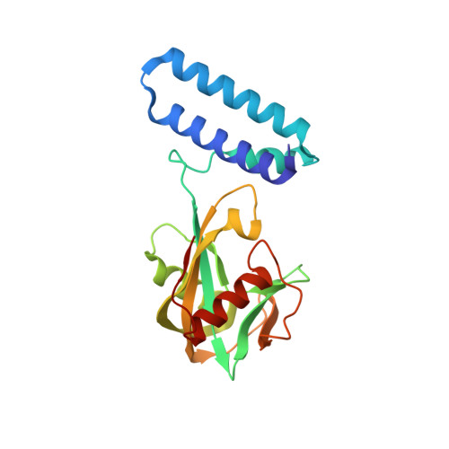
    4HFQ

  • PubMed Abstract: 

    The gene for a Nudix enzyme (SP_1669) was found to code for a UDP-X diphosphatase. The SP_1669 gene is localized among genes encoding proteins that participate in cell division in Streptococcus pneumoniae. One of these genes, MurF, encodes an enzyme that catalyzes the last step of the Mur pathway of peptidoglycan biosynthesis. Mur pathway substrates are all derived from UDP-glucosamine and all are potential Nudix substrates. We showed that UDP-X diphosphatase can hydrolyze the Mur pathway substrates UDP-N-acetylmuramic acid and UDP-N-acetylmuramoyl-L-alanine. The 1.39 Å resolution crystal structure of this enzyme shows that it folds as an asymmetric homodimer with two distinct active sites, each containing elements of the conserved Nudix box sequence. In addition to its Nudix catalytic activity, the enzyme has a 3'5' RNA exonuclease activity. We propose that the structural asymmetry in UDP-X diphosphatase facilitates the recognition of these two distinct classes of substrates, Nudix substrates and RNA. UDP-X diphosphatase is a prototype of a new family of Nudix enzymes with unique structural characteristics: two monomers, each consisting of an N-terminal helix bundle domain and a C-terminal Nudix domain, form an asymmetric dimer with two distinct active sites. These enzymes function to hydrolyze bacterial cell wall precursors and degrade RNA.


  • Organizational Affiliation
    • Department of Biophysics and Biophysical Chemistry, Johns Hopkins University School of Medicine, Baltimore, Maryland, United States of America.

Macromolecule Content 

  • Total Structure Weight: 49.64 kDa 
  • Atom Count: 4,205 
  • Modeled Residue Count: 406 
  • Deposited Residue Count: 406 
  • Unique protein chains: 1

Macromolecules

Find similar proteins by:|  3D Structure
Entity ID: 1
MoleculeChains  Sequence LengthOrganismDetailsImage
MutT/nudix family protein
A, B
203Streptococcus pneumoniae TIGR4Mutation(s): 0 
Gene Names: SP1669SP_1669
UniProt
Find proteins for A0A0H2URA0 (Streptococcus pneumoniae serotype 4 (strain ATCC BAA-334 / TIGR4))
Explore A0A0H2URA0 
Go to UniProtKB:  A0A0H2URA0
Entity Groups
Sequence Clusters30% Identity50% Identity70% Identity90% Identity95% Identity100% Identity
UniProt GroupA0A0H2URA0
Sequence Annotations
Expand
Reference Sequence

Small Molecules

Ligands 6 Unique
IDChains Name / Formula / InChI Key2D Diagram3D Interactions
PGE

Query on PGE


Download:Ideal Coordinates CCD File
O [auth A]TRIETHYLENE GLYCOL
C6 H14 O4
ZIBGPFATKBEMQZ-UHFFFAOYSA-N
TRS

Query on TRS


Download:Ideal Coordinates CCD File
E [auth A],
X [auth B],
Z [auth B]
2-AMINO-2-HYDROXYMETHYL-PROPANE-1,3-DIOL
C4 H12 N O3
LENZDBCJOHFCAS-UHFFFAOYSA-O
PEG

Query on PEG


Download:Ideal Coordinates CCD File
FA [auth B],
I [auth A],
L [auth A],
R [auth A]
DI(HYDROXYETHYL)ETHER
C4 H10 O3
MTHSVFCYNBDYFN-UHFFFAOYSA-N
SO4

Query on SO4


Download:Ideal Coordinates CCD File
F [auth A],
G [auth A],
H [auth A],
M [auth A],
T [auth B]
SULFATE ION
O4 S
QAOWNCQODCNURD-UHFFFAOYSA-L
GOL

Query on GOL


Download:Ideal Coordinates CCD File
AA [auth B]
K [auth A]
P [auth A]
Q [auth A]
V [auth B]
AA [auth B],
K [auth A],
P [auth A],
Q [auth A],
V [auth B],
W [auth B]
GLYCEROL
C3 H8 O3
PEDCQBHIVMGVHV-UHFFFAOYSA-N
EDO

Query on EDO


Download:Ideal Coordinates CCD File
BA [auth B]
C [auth A]
CA [auth B]
D [auth A]
DA [auth B]
BA [auth B],
C [auth A],
CA [auth B],
D [auth A],
DA [auth B],
EA [auth B],
J [auth A],
N [auth A],
S [auth A],
U [auth B],
Y [auth B]
1,2-ETHANEDIOL
C2 H6 O2
LYCAIKOWRPUZTN-UHFFFAOYSA-N

Experimental Data & Validation

Experimental Data

  • Method: X-RAY DIFFRACTION
  • Resolution: 1.39 Å
  • R-Value Free:  0.196 (Depositor), 0.195 (DCC) 
  • R-Value Work:  0.172 (Depositor), 0.171 (DCC) 
  • R-Value Observed: 0.174 (Depositor) 
Space Group: P 31
Unit Cell:
Length ( Å )Angle ( ˚ )
a = 77.992α = 90
b = 77.992β = 90
c = 64.704γ = 120
Software Package:
Software NamePurpose
DENZOdata reduction
SCALEPACKdata scaling
SOLVEphasing
REFMACrefinement
PDB_EXTRACTdata extraction

Structure Validation

View Full Validation Report


Entry History 

Deposition Data

Revision History  (Full details and data files)

  • Version 1.0: 2013-08-07
    Type: Initial release
  • Version 1.1: 2024-02-28
    Changes: Data collection, Database references, Derived calculations