4HCO

Human Plk1-PBD in complex with Thymoquinone at the phophopeptide binding site


Experimental Data Snapshot

  • Method: X-RAY DIFFRACTION
  • Resolution: 2.75 Å
  • R-Value Free: 0.280 
  • R-Value Work: 0.206 
  • R-Value Observed: 0.210 

wwPDB Validation   3D Report Full Report


Ligand Structure Quality Assessment 


This is version 1.2 of the entry. See complete history


Literature

Thymoquinone Blocks pSer/pThr Recognition by Plk1 Polo-Box Domain As a Phosphate Mimic

Yin, Z.Song, Y.Rehse, P.H.

(2013) ACS Chem Biol 8: 303-308

  • DOI: https://doi.org/10.1021/cb3004379
  • Primary Citation of Related Structures:  
    4H5X, 4H71, 4HCO

  • PubMed Abstract: 

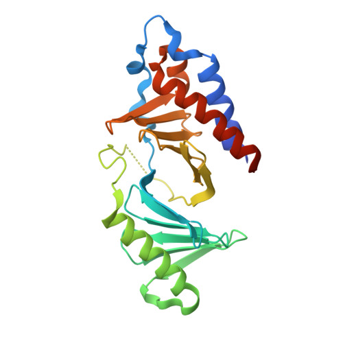
    Phosphorylation-dependent protein-protein interaction has rarely been targeted in medicinal chemistry. Thymoquinone, a naturally occurring antitumor agent, disrupts prephosphorylated substrate recognition by the polo-box domain of polo-like kinase 1, a key mitotic regulator responsible for various carcinogenesis when overexpressed. Here, crystallographic studies reveal that the phosphoserine/phosphothreonine recognition site of the polo-box domain is the binding pocket for thymoquinone and its analogue poloxime. Both small molecules displace phosphopeptides bound with the polo-box domain in a slow but noncovalent binding mode. A conserved water bridge and a cation-π interaction were found as their competition strategy against the phosphate group. This mechanism sheds light on small-molecule intervention of phospho-recognition by the polo-box domain of polo-like kinase 1 and other phospho-binding proteins in general.


  • Organizational Affiliation

    School of Life Science and Technology, China Pharmaceutical University, 24 Tong Jia Xiang, Nanjing, Jiangsu 210009, China.


Macromolecules
Find similar proteins by:  (by identity cutoff)  |  3D Structure
Entity ID: 1
MoleculeChains Sequence LengthOrganismDetailsImage
Serine/threonine-protein kinase PLK1A [auth B],
B [auth A]
240Homo sapiensMutation(s): 0 
Gene Names: PLK1PLK
EC: 2.7.11.21
UniProt & NIH Common Fund Data Resources
Find proteins for P53350 (Homo sapiens)
Explore P53350 
Go to UniProtKB:  P53350
PHAROS:  P53350
GTEx:  ENSG00000166851 
Entity Groups  
Sequence Clusters30% Identity50% Identity70% Identity90% Identity95% Identity100% Identity
UniProt GroupP53350
Sequence Annotations
Expand
  • Reference Sequence
Small Molecules
Ligands 2 Unique
IDChains Name / Formula / InChI Key2D Diagram3D Interactions
IMW
Query on IMW

Download Ideal Coordinates CCD File 
D [auth A]2-methyl-5-(1-methylethyl)cyclohexa-2,5-diene-1,4-dione
C10 H12 O2
KEQHJBNSCLWCAE-UHFFFAOYSA-N
GOL
Query on GOL

Download Ideal Coordinates CCD File 
C [auth A]GLYCEROL
C3 H8 O3
PEDCQBHIVMGVHV-UHFFFAOYSA-N
Binding Affinity Annotations 
IDSourceBinding Affinity
IMW PDBBind:  4HCO IC50: 4.74e+4 (nM) from 1 assay(s)
BindingDB:  4HCO IC50: 2180 (nM) from 1 assay(s)
Experimental Data & Validation

Experimental Data

  • Method: X-RAY DIFFRACTION
  • Resolution: 2.75 Å
  • R-Value Free: 0.280 
  • R-Value Work: 0.206 
  • R-Value Observed: 0.210 
  • Space Group: P 1 21 1
Unit Cell:
Length ( Å )Angle ( ˚ )
a = 33.26α = 90
b = 102.15β = 93.96
c = 67.74γ = 90
Software Package:
Software NamePurpose
MAR345dtbdata collection
MOLREPphasing
REFMACrefinement
MOSFLMdata reduction
SCALAdata scaling

Structure Validation

View Full Validation Report


Ligand Structure Quality Assessment 


Entry History 

Deposition Data

Revision History  (Full details and data files)

  • Version 1.0: 2012-10-10
    Type: Initial release
  • Version 1.1: 2013-03-06
    Changes: Database references
  • Version 1.2: 2023-11-08
    Changes: Data collection, Database references, Derived calculations, Refinement description, Structure summary