4HCA | pdb_00004hca

DNA binding by GATA transcription factor-complex 1


Experimental Data Snapshot

  • Method: X-RAY DIFFRACTION
  • Resolution: 2.80 Å
  • R-Value Free: 
    0.296 (Depositor), 0.284 (DCC) 
  • R-Value Work: 
    0.245 (Depositor), 0.251 (DCC) 
  • R-Value Observed: 
    0.247 (Depositor) 

wwPDB Validation 3D Report Full Report

Validation slider image for 4HCA

This is version 1.2 of the entry. See complete history

Literature

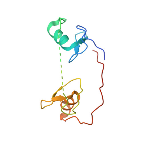
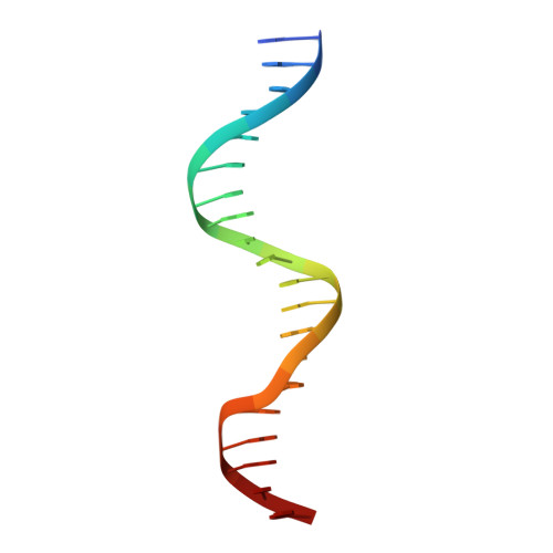
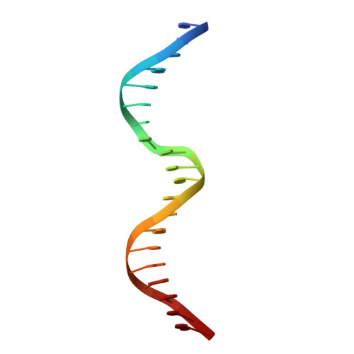
DNA Binding by GATA Transcription Factor Suggests Mechanisms of DNA Looping and Long-Range Gene Regulation.

Chen, Y.Bates, D.L.Dey, R.Chen, P.H.Machado, A.C.Laird-Offringa, I.A.Rohs, R.Chen, L.

(2012) Cell Rep 2: 1197-1206

  • DOI: https://doi.org/10.1016/j.celrep.2012.10.012
  • Primary Citation Related Structures: 
    4HC7, 4HC9, 4HCA

  • PubMed Abstract: 

    GATA transcription factors regulate transcription during development and differentiation by recognizing distinct GATA sites with a tandem of two conserved zinc fingers, and by mediating long-range DNA looping. However, the molecular basis of these processes is not well understood. Here, we determined three crystal structures of the full DNA-binding domain (DBD) of human GATA3 protein, which contains both zinc fingers, in complex with different DNA sites. In one structure, both zinc fingers wrap around a palindromic GATA site, cooperatively enhancing the binding affinity and kinetic stability. Strikingly, in the other two structures, the two fingers of GATA DBD bind GATA sites on different DNA molecules, thereby bridging two separate DNA fragments. This was confirmed in solution by an in-gel fluorescence resonance energy transfer analysis. These findings not only provide insights into the structure and function of GATA proteins but also shed light on the molecular basis of long-range gene regulation.


  • Organizational Affiliation
    • Molecular and Computational Biology Program, Departments of Biological Sciences and Chemistry, University of Southern California, Los Angeles, CA 90089, USA.

Macromolecule Content 

  • Total Structure Weight: 25.29 kDa 
  • Atom Count: 1,591 
  • Modeled Residue Count: 139 
  • Deposited Residue Count: 155 
  • Unique protein chains: 1
  • Unique nucleic acid chains: 2

Macromolecules


Find similar proteins by:|  3D Structure
Entity ID: 1
MoleculeChains  Sequence LengthOrganismDetailsImage
Trans-acting T-cell-specific transcription factor GATA-3115Homo sapiensMutation(s): 0 
Gene Names: GATA3
UniProt & NIH Common Fund Data Resources
Find proteins for P23771 (Homo sapiens)
Explore P23771 
Go to UniProtKB:  P23771
PHAROS:  P23771
GTEx:  ENSG00000107485 
Entity Groups
Sequence Clusters30% Identity50% Identity70% Identity90% Identity95% Identity100% Identity
UniProt GroupP23771
Sequence Annotations
Expand
Reference Sequence
Find similar nucleic acids by:  Sequence
Entity ID: 2
MoleculeChains LengthOrganismImage
DNA (5'-D(*AP*AP*TP*GP*TP*CP*CP*AP*TP*CP*TP*GP*AP*TP*AP*AP*GP*AP*CP*G)-3')B [auth X]20N/A
Sequence Annotations
Expand
Reference Sequence
Find similar nucleic acids by:  Sequence
Entity ID: 3
MoleculeChains LengthOrganismImage
DNA (5'-D(*TP*TP*CP*GP*TP*CP*TP*TP*AP*TP*CP*AP*GP*AP*TP*GP*GP*AP*CP*A)-3')C [auth Y]20N/A
Sequence Annotations
Expand
Reference Sequence

Experimental Data & Validation

Experimental Data

  • Method: X-RAY DIFFRACTION
  • Resolution: 2.80 Å
  • R-Value Free:  0.296 (Depositor), 0.284 (DCC) 
  • R-Value Work:  0.245 (Depositor), 0.251 (DCC) 
  • R-Value Observed: 0.247 (Depositor) 
Space Group: P 1 21 1
Unit Cell:
Length ( Å )Angle ( ˚ )
a = 54.486α = 90
b = 36.55β = 103.7
c = 66.118γ = 90
Software Package:
Software NamePurpose
PHENIXrefinement
PDB_EXTRACTdata extraction

Structure Validation

View Full Validation Report


Entry History 

Deposition Data

Revision History  (Full details and data files)

  • Version 1.0: 2012-12-05
    Type: Initial release
  • Version 1.1: 2013-01-02
    Changes: Database references
  • Version 1.2: 2024-02-28
    Changes: Data collection, Database references, Derived calculations