4HA6

Crystal structure of pyridoxine 4-oxidase - pyridoxamine complex


Experimental Data Snapshot

  • Method: X-RAY DIFFRACTION
  • Resolution: 2.10 Å
  • R-Value Free: 0.233 
  • R-Value Work: 0.186 
  • R-Value Observed: 0.188 

wwPDB Validation   3D Report Full Report


Ligand Structure Quality Assessment 


This is version 1.1 of the entry. See complete history


Literature

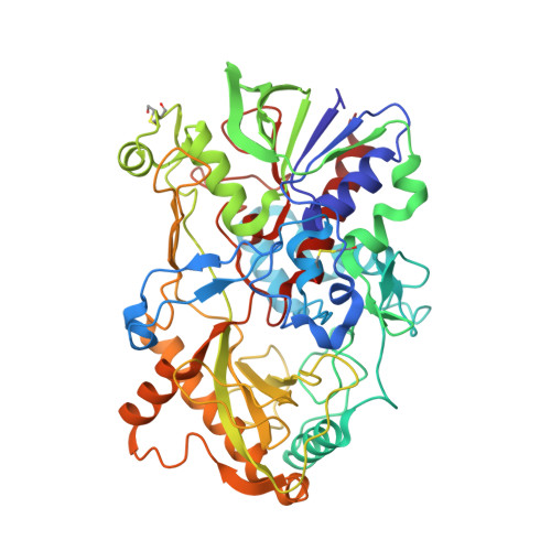
Crystal structure of pyridoxine 4-oxidase from Mesorhizobium loti.

Mugo, A.N.Kobayashi, J.Yamasaki, T.Mikami, B.Ohnishi, K.Yoshikane, Y.Yagi, T.

(2013) Biochim Biophys Acta 1834: 953-963

  • DOI: https://doi.org/10.1016/j.bbapap.2013.03.004
  • Primary Citation of Related Structures:  
    4HA6

  • PubMed Abstract: 

    Pyridoxine 4-oxidase (PNOX) from Mesorhizobium loti is a monomeric glucose-methanol-choline (GMC) oxidoreductase family enzyme, catalyzes FAD-dependent oxidation of pyridoxine (PN) into pyridoxal, and is the first enzyme in pathway I for the degradation of PN. The tertiary structures of PNOX with a C-terminal His6-tag and PNOX-pyridoxamine (PM) complex were determined at 2.2Å and at 2.1Å resolutions, respectively. The overall structure consisted of FAD-binding and substrate-binding domains. In the active site, His460, His462, and Pro504 were located on the re-face of the isoalloxazine ring of FAD. PM binds to the active site through several hydrogen bonds. The side chains of His462 and His460 are located at 2.7 and 3.1Å from the N4' atom of PM. The activities of His460Ala and His462Ala mutant PNOXs were very low, and 460Ala/His462Ala double mutant PNOX exhibited no activity. His462 may act as a general base for the abstraction of a proton from the 4'-hydroxyl of PN. His460 may play a role in the binding and positioning of PN. The C4' atom in PM is located at 3.2Å, and the hydride ion from the C4' atom may be transferred to the N5 atom of the isoalloxazine ring. The comparison of active site residues in GMC oxidoreductase shows that Pro504 in PNOX corresponds to Asn or His of the conserved His-Asn or His-His pair in other GMC oxidoreductases. The function of the novel proline residue was discussed.


  • Organizational Affiliation

    Graduate School of Integral Arts and Science, Kochi University, Nankoku, Kochi, Japan.


Macromolecules
Find similar proteins by:  (by identity cutoff)  |  3D Structure
Entity ID: 1
MoleculeChains Sequence LengthOrganismDetailsImage
Pyridoxine 4-oxidase508Mesorhizobium lotiMutation(s): 0 
Gene Names: mll6785pno
EC: 1.1.3.12
UniProt
Find proteins for Q5NT46 (Rhizobium loti)
Explore Q5NT46 
Go to UniProtKB:  Q5NT46
Entity Groups  
Sequence Clusters30% Identity50% Identity70% Identity90% Identity95% Identity100% Identity
UniProt GroupQ5NT46
Sequence Annotations
Expand
  • Reference Sequence
Small Molecules
Ligands 3 Unique
IDChains Name / Formula / InChI Key2D Diagram3D Interactions
FAD
Query on FAD

Download Ideal Coordinates CCD File 
B [auth A]FLAVIN-ADENINE DINUCLEOTIDE
C27 H33 N9 O15 P2
VWWQXMAJTJZDQX-UYBVJOGSSA-N
PXM
Query on PXM

Download Ideal Coordinates CCD File 
J [auth A]4-(AMINOMETHYL)-5-(HYDROXYMETHYL)-2-METHYLPYRIDIN-3-OL
C8 H12 N2 O2
NHZMQXZHNVQTQA-UHFFFAOYSA-N
GOL
Query on GOL

Download Ideal Coordinates CCD File 
C [auth A]
D [auth A]
E [auth A]
F [auth A]
G [auth A]
C [auth A],
D [auth A],
E [auth A],
F [auth A],
G [auth A],
H [auth A],
I [auth A]
GLYCEROL
C3 H8 O3
PEDCQBHIVMGVHV-UHFFFAOYSA-N
Modified Residues  1 Unique
IDChains TypeFormula2D DiagramParent
CME
Query on CME
A
L-PEPTIDE LINKINGC5 H11 N O3 S2CYS
Experimental Data & Validation

Experimental Data

  • Method: X-RAY DIFFRACTION
  • Resolution: 2.10 Å
  • R-Value Free: 0.233 
  • R-Value Work: 0.186 
  • R-Value Observed: 0.188 
  • Space Group: P 21 21 21
Unit Cell:
Length ( Å )Angle ( ˚ )
a = 62.489α = 90
b = 79.169β = 90
c = 135.219γ = 90
Software Package:
Software NamePurpose
PHENIXrefinement
PDB_EXTRACTdata extraction
HKL-2000data collection
HKL-2000data reduction
HKL-2000data scaling
CCP4phasing

Structure Validation

View Full Validation Report


Ligand Structure Quality Assessment 


Entry History 

Deposition Data

Revision History  (Full details and data files)

  • Version 1.0: 2013-06-05
    Type: Initial release
  • Version 1.1: 2023-11-08
    Changes: Data collection, Database references, Derived calculations, Refinement description