The Near-iron Transporter (NEAT) Domains of the Anthrax Hemophore IsdX2 Require a Critical Glutamine to Extract Heme from Methemoglobin.
Honsa, E.S., Owens, C.P., Goulding, C.W., Maresso, A.W.(2013) J Biological Chem 288: 8479-8490
- PubMed: 23364793 Search on PubMedSearch on PubMed Central
- DOI: https://doi.org/10.1074/jbc.M112.430009
- Primary Citation Related Structures:
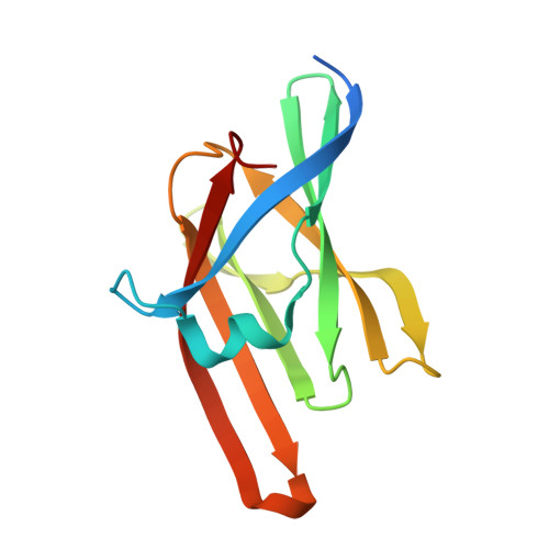
4H8P, 4H8Q - PubMed Abstract:
Several gram-positive pathogenic bacteria employ near-iron transporter (NEAT) domains to acquire heme from hemoglobin during infection. However, the structural requirements and mechanism of action for NEAT-mediated heme extraction remains unknown. Bacillus anthracis exhibits a rapid growth rate during systemic infection, suggesting that the bacterium expresses efficient iron acquisition systems. To understand how B. anthracis acquires iron from heme sources, which account for 80% of mammalian iron stores, we investigated the properties of the five-NEAT domain hemophore IsdX2. Using a combination of bioinformatics and site-directed mutagenesis, we determined that the heme extraction properties of IsdX2 are dependent on an amino acid with an amide side chain within the 310-helix of the NEAT domain. Additionally, we used a spectroscopic analysis to show that IsdX2 NEAT domains only scavenge heme from methemoglobin (metHb) and that autoxidation of oxyhemoglobin to metHb must occur prior to extraction. We also report the crystal structures of NEAT5 wild type and a Q29T mutant and present surface plasmon resonance data that indicate that the loss of this amide side chain reduces the affinity of the NEAT domain for metHb. We propose a model whereby the amide side chain is first required to drive an interaction with metHb that destabilizes heme, which is subsequently extracted and coordinated in the aliphatic heme-binding environment of the NEAT domain. Because an amino acid with an amide side chain in this position is observed in NEAT domains of several genera of gram-positive pathogenic bacteria, these results suggest that specific targeting of this or nearby residues may be an entry point for inhibitor development aimed at blocking bacterial iron acquisition during infection.
- Department of Molecular Virology and Microbiology, Baylor College of Medicine, Houston, Texas 77030.
Organizational Affiliation: