


Crystal structure of a putative alpha-L-fucosidase (BT_0435) from Bacteroides thetaiotaomicron VPI-5482 at 1.80 A resolution
Joint Center for Structural Genomics (JCSG)To be published.
Experimental Data Snapshot
wwPDB Validation 3D Report Full Report

Entity ID: 1 | |||||
|---|---|---|---|---|---|
| Molecule | Chains | Sequence Length | Organism | Details | Image |
| putative alpha-L-fucosidase | 340 | Bacteroides thetaiotaomicron VPI-5482 | Mutation(s): 0 Gene Names: BT_0435, NP_809348.1 | 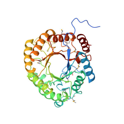 | |
UniProt | |||||
Entity Groups | |||||
| Sequence Clusters | 30% Identity50% Identity70% Identity90% Identity95% Identity100% Identity | ||||
| UniProt Group | Q8AAM9 | ||||
Sequence AnnotationsExpand | |||||
Reference Sequence | |||||
| Ligands 6 Unique | |||||
|---|---|---|---|---|---|
| ID | Chains | Name / Formula / InChI Key | 2D Diagram | 3D Interactions | |
| PE4 Download:Ideal Coordinates CCD File | G [auth A], H [auth A] | 2-{2-[2-(2-{2-[2-(2-ETHOXY-ETHOXY)-ETHOXY]-ETHOXY}-ETHOXY)-ETHOXY]-ETHOXY}-ETHANOL C16 H34 O8 PJWQOENWHPEPKI-UHFFFAOYSA-N |  | ||
| PG6 Download:Ideal Coordinates CCD File | O [auth B], P [auth B] | 1-(2-METHOXY-ETHOXY)-2-{2-[2-(2-METHOXY-ETHOXY]-ETHOXY}-ETHANE C12 H26 O6 DMDPGPKXQDIQQG-UHFFFAOYSA-N |  | ||
| 1PE Download:Ideal Coordinates CCD File | E [auth A] F [auth A] I [auth A] N [auth B] R [auth B] | PENTAETHYLENE GLYCOL C10 H22 O6 JLFNLZLINWHATN-UHFFFAOYSA-N |  | ||
| PG4 Download:Ideal Coordinates CCD File | J [auth A], K [auth A], L [auth A], S [auth B], W [auth C] | TETRAETHYLENE GLYCOL C8 H18 O5 UWHCKJMYHZGTIT-UHFFFAOYSA-N |  | ||
| NA Download:Ideal Coordinates CCD File | Q [auth B] | SODIUM ION Na FKNQFGJONOIPTF-UHFFFAOYSA-N |  | ||
| UNL Download:Ideal Coordinates CCD File | D [auth A], M [auth B], T [auth C] | Unknown ligand FKNQFGJONOIPTF-UHFFFAOYSA-N | |||
| Modified Residues 1 Unique | |||||
|---|---|---|---|---|---|
| ID | Chains | Type | Formula | 2D Diagram | Parent |
| MSE Query on MSE | A, B, C | L-PEPTIDE LINKING | C5 H11 N O2 Se |  | MET |
| Length ( Å ) | Angle ( ˚ ) |
|---|---|
| a = 53.67 | α = 90 |
| b = 125.7 | β = 103.72 |
| c = 80.01 | γ = 90 |
| Software Name | Purpose |
|---|---|
| MolProbity | model building |
| PDB_EXTRACT | data extraction |
| SHELX | phasing |
| SHARP | phasing |
| SCALA | data scaling |
| BUSTER-TNT | refinement |
| MOSFLM | data reduction |
| SHELXD | phasing |
| BUSTER | refinement |


