4H33 | pdb_00004h33

Crystal structure of a voltage-gated K+ channel pore module in a closed state in lipid membranes, tetragonal crystal form


Experimental Data Snapshot

  • Method: X-RAY DIFFRACTION
  • Resolution: 3.10 Å
  • R-Value Free: 
    0.305 (Depositor), 0.330 (DCC) 
  • R-Value Work: 
    0.281 (Depositor), 0.310 (DCC) 
  • R-Value Observed: 
    0.284 (Depositor) 

Starting Models: experimental
View more details

wwPDB Validation 3D Report Full Report

Validation slider image for 4H33

Ligand Structure Quality Assessment 


This is version 1.3 of the entry. See complete history

Literature

Crystal Structure of a Voltage-gated K+ Channel Pore Module in a Closed State in Lipid Membranes.

Santos, J.S.Asmar-Rovira, G.A.Han, G.W.Liu, W.Syeda, R.Cherezov, V.Baker, K.A.Stevens, R.C.Montal, M.

(2012) J Biological Chem 287: 43063-43070

  • DOI: https://doi.org/10.1074/jbc.M112.415091
  • Primary Citation Related Structures: 
    4H33, 4H37

  • PubMed Abstract: 

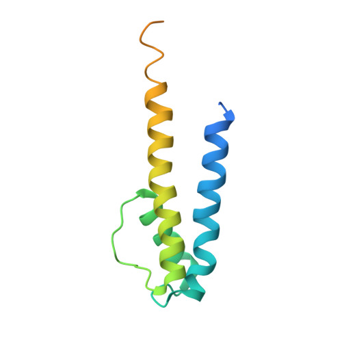
    Voltage-gated K(+) channels underlie the electrical excitability of cells. Each subunit of the functional tetramer consists of the tandem fusion of two modules, an N-terminal voltage-sensor and a C-terminal pore. To investigate how sensor coupling to the pore generates voltage-dependent channel opening, we solved the crystal structure and characterized the function of a voltage-gated K(+) channel pore in a lipid membrane. The structure of a functional channel in a membrane environment at 3.1 Å resolution establishes an unprecedented connection between channel structure and function. The structure is unique in delineating an ion-occupied ready to conduct selectivity filter, a confined aqueous cavity, and a closed activation gate, embodying a dynamic entity trapped in an unstable closed state.


  • Organizational Affiliation
    • Section of Neurobiology, Division of Biological Sciences, University of California San Diego, La Jolla, California 92093, USA.

Macromolecule Content 

  • Total Structure Weight: 17.1 kDa 
  • Atom Count: 733 
  • Modeled Residue Count: 91 
  • Deposited Residue Count: 137 
  • Unique protein chains: 1

Macromolecules

Find similar proteins by:|  3D Structure
Entity ID: 1
MoleculeChains  Sequence LengthOrganismDetailsImage
Lmo2059 protein137Listeria monocytogenes EGD-eMutation(s): 1 
Gene Names: lmo2059
Membrane Entity: Yes 
UniProt
Find proteins for Q8Y5K1 (Listeria monocytogenes serovar 1/2a (strain ATCC BAA-679 / EGD-e))
Explore Q8Y5K1 
Go to UniProtKB:  Q8Y5K1
Entity Groups
Sequence Clusters30% Identity50% Identity70% Identity90% Identity95% Identity100% Identity
UniProt GroupQ8Y5K1
Sequence Annotations
Expand
Reference Sequence

Experimental Data & Validation

Experimental Data

  • Method: X-RAY DIFFRACTION
  • Resolution: 3.10 Å
  • R-Value Free:  0.305 (Depositor), 0.330 (DCC) 
  • R-Value Work:  0.281 (Depositor), 0.310 (DCC) 
  • R-Value Observed: 0.284 (Depositor) 
Space Group: P 4 21 2
Unit Cell:
Length ( Å )Angle ( ˚ )
a = 53.37α = 90
b = 53.37β = 90
c = 108.16γ = 90
Software Package:
Software NamePurpose
Blu-Icedata collection
PHASERphasing
BUSTERrefinement
HKL-2000data reduction
HKL-2000data scaling

Structure Validation

View Full Validation Report


Ligand Structure Quality Assessment 


Entry History 

Deposition Data

Revision History  (Full details and data files)

  • Version 1.0: 2012-11-07
    Type: Initial release
  • Version 1.1: 2013-01-16
    Changes: Database references
  • Version 1.2: 2017-11-15
    Changes: Refinement description
  • Version 1.3: 2023-09-20
    Changes: Data collection, Database references, Derived calculations, Refinement description