Crystal Structure of a Promoter Sequence in the B-raf Gene Reveals an Intertwined Dimer Quadruplex.
Wei, D., Todd, A.K., Zloh, M., Gunaratnam, M., Parkinson, G.N., Neidle, S.(2013) J Am Chem Soc 135: 19319-19329
- PubMed: 24295054 Search on PubMed
- DOI: https://doi.org/10.1021/ja4101358
- Primary Citation Related Structures:
4H29 - PubMed Abstract:
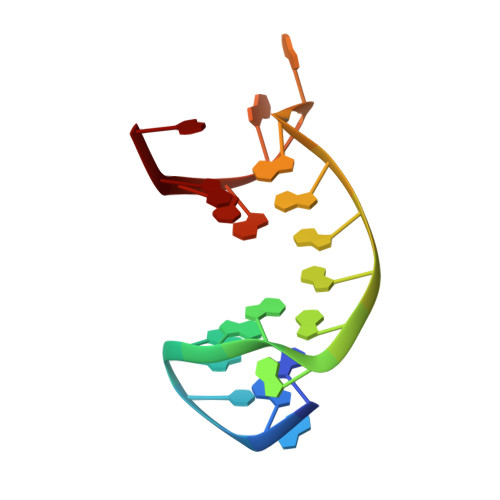
The sequence d(GGGCGGGGAGGGGGAAGGGA) occurs in the promoter region of the B-raf gene. An X-ray crystallographic study has found that this forms an unprecedented dimeric quadruplex arrangement, with a core of seven consecutive G-quartets and an uninterrupted run of six potassium ions in the central channel of the quadruplex. Analogy with previously reported promoter quadruplexes had initially suggested that in common with these a monomeric quadruplex was to be expected. The structure has a distorted G·C·G·C base quartet at one end and four flipped-out adenosine nucleosides at the other. The only loops in the structure are formed by the cytosine and by the three adenosines within the sequence, with all of the guanosines participating in G-quartet formation. Solution UV and circular dichroism data are in accord with a stable quadruple arrangement being formed. 1D NMR data, together with gel electrophoresis measurements, are consistent with a dimer being the dominant species in potassium solution. A single-chain intramolecular quadruplex has been straightforwardly constructed using molecular modeling, by means of a six-nucleotide sequence joining 3' and 5' ends of each strand in the dimer. A human genomic database search has revealed a number of sequences containing eight or more consecutive short G-tracts, suggesting that such intramolecular quadruplexes could be formed within the human genome.
- UCL School of Pharmacy, University College London , London WC1N 1AX, United Kingdom.
Organizational Affiliation: