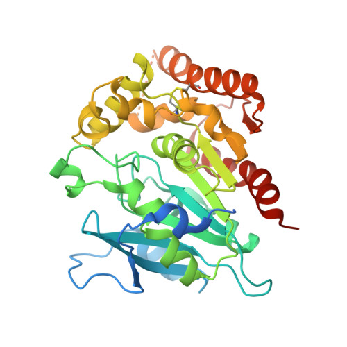
4H18

Three dimensional structure of corynomycoloyl tranferase C


Experimental Data Snapshot

  • Method: X-RAY DIFFRACTION
  • Resolution: 1.75 Å
  • R-Value Free: 0.203 
  • R-Value Work: 0.163 
  • R-Value Observed: 0.165 

Starting Model: experimental
View more details

wwPDB Validation   3D Report Full Report


This is version 1.2 of the entry. See complete history


Literature

Identification of a mycoloyl transferase selectively involved in o-acylation of polypeptides in corynebacteriales.

Huc, E.de Sousa-D'Auria, C.de la Sierra-Gallay, I.L.Salmeron, C.van Tilbeurgh, H.Bayan, N.Houssin, C.Daffe, M.Tropis, M.

(2013) J Bacteriol 195: 4121-4128

  • DOI: https://doi.org/10.1128/JB.00285-13
  • Primary Citation of Related Structures:  
    4H18

  • PubMed Abstract: 

    We have previously described the posttranslational modification of pore-forming small proteins of Corynebacterium by mycolic acid, a very-long-chain α-alkyl and β-hydroxy fatty acid. Using a combination of chemical analyses and mass spectrometry, we identified the mycoloyl transferase (Myt) that catalyzes the transfer of the fatty acid residue to yield O-acylated polypeptides. Inactivation of corynomycoloyl transferase C (cg0413 [Corynebacterium glutamicum mytC {CgmytC}]), one of the six Cgmyt genes of C. glutamicum, specifically abolished the O-modification of the pore-forming proteins PorA and PorH, which is critical for their biological activity. Expectedly, complementation of the cg0413 mutant with either the wild-type gene or its orthologues from Corynebacterium diphtheriae and Rhodococcus, but not Nocardia, fully restored the O-acylation of the porins. Consistently, the three-dimensional structure of CgMytC showed the presence of a unique loop that is absent from enzymes that transfer mycoloyl residues onto both trehalose and the cell wall arabinogalactan. These data suggest the implication of this structure in the enzyme specificity for protein instead of carbohydrate.


  • Organizational Affiliation

    Team Enveloppes Mycobactériennes, Structure Biosynthèse et Rôles, Centre National de la Recherche Scientifique (CNRS), Institut de Pharmacologie et Biologie Structurale (IPBS), Département Mécanismes Moléculaires des Infections Mycobactériennes, UMR 5089, Toulouse, France.


Macromolecules
Find similar proteins by:  (by identity cutoff)  |  3D Structure
Entity ID: 1
MoleculeChains Sequence LengthOrganismDetailsImage
Cmt1
A, B, C, D
371Corynebacterium glutamicum ATCC 13032Mutation(s): 0 
Gene Names: cmt1Cmt1cg0413Cgl0343WA5_0336
EC: 2.3.1.122
UniProt
Find proteins for Q8NTG4 (Corynebacterium glutamicum (strain ATCC 13032 / DSM 20300 / JCM 1318 / BCRC 11384 / CCUG 27702 / LMG 3730 / NBRC 12168 / NCIMB 10025 / NRRL B-2784 / 534))
Explore Q8NTG4 
Go to UniProtKB:  Q8NTG4
Entity Groups  
Sequence Clusters30% Identity50% Identity70% Identity90% Identity95% Identity100% Identity
UniProt GroupQ8NTG4
Sequence Annotations
Expand
  • Reference Sequence
Small Molecules
Ligands 1 Unique
IDChains Name / Formula / InChI Key2D Diagram3D Interactions
MG
Query on MG

Download Ideal Coordinates CCD File 
E [auth D]MAGNESIUM ION
Mg
JLVVSXFLKOJNIY-UHFFFAOYSA-N
Experimental Data & Validation

Experimental Data

  • Method: X-RAY DIFFRACTION
  • Resolution: 1.75 Å
  • R-Value Free: 0.203 
  • R-Value Work: 0.163 
  • R-Value Observed: 0.165 
  • Space Group: P 21 21 2
Unit Cell:
Length ( Å )Angle ( ˚ )
a = 85.804α = 90
b = 190.688β = 90
c = 78.497γ = 90
Software Package:
Software NamePurpose
ADSCdata collection
PHASERphasing
PHENIXrefinement
XDSdata reduction
XDSdata scaling

Structure Validation

View Full Validation Report


Entry History 

Revision History  (Full details and data files)

  • Version 1.0: 2013-09-25
    Type: Initial release
  • Version 1.1: 2018-01-24
    Changes: Structure summary
  • Version 1.2: 2023-09-13
    Changes: Data collection, Database references, Derived calculations, Refinement description