4GYV | pdb_00004gyv

Crystal structure of the DH domain of FARP2


Experimental Data Snapshot

  • Method: X-RAY DIFFRACTION
  • Resolution: 2.90 Å
  • R-Value Free: 
    0.221 (Depositor), 0.229 (DCC) 
  • R-Value Work: 
    0.178 (Depositor), 0.182 (DCC) 
  • R-Value Observed: 
    0.179 (Depositor) 

wwPDB Validation 3D Report Full Report

Validation slider image for 4GYV

This is version 1.4 of the entry. See complete history

Literature

Structural Basis for Autoinhibition of the Guanine Nucleotide Exchange Factor FARP2.

He, X.Kuo, Y.C.Rosche, T.J.Zhang, X.

(2013) Structure 21: 355-364

  • DOI: https://doi.org/10.1016/j.str.2013.01.001
  • Primary Citation Related Structures: 
    4GYV, 4GZU, 4H6Y

  • PubMed Abstract: 

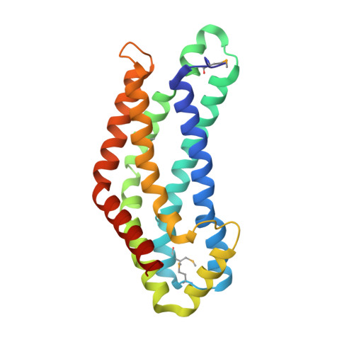
    FARP2 is a Dbl-family guanine nucleotide exchange factor (GEF) that contains a 4.1, ezrin, radixin and moesin (FERM) domain, a Dbl-homology (DH) domain and two pleckstrin homology (PH) domains. FARP2 activates Rac1 or Cdc42 in response to upstream signals, thereby regulating processes such as neuronal axon guidance and bone homeostasis. How the GEF activity of FARP2 is regulated remained poorly understood. We have determined the crystal structures of the catalytic DH domain and the DH-PH-PH domains of FARP2. The structures reveal an auto-inhibited conformation in which the GEF substrate-binding site is blocked collectively by the last helix in the DH domain and the two PH domains. This conformation is stabilized by multiple interactions among the domains and two well-structured inter-domain linkers. Our cell-based activity assays confirm the suppression of the FARP2 GEF activity by these auto-inhibitory elements.


  • Organizational Affiliation
    • Department of Pharmacology, University of Texas Southwestern Medical Center, Dallas, TX 75063, USA.

Macromolecule Content 

  • Total Structure Weight: 232.11 kDa 
  • Atom Count: 15,733 
  • Modeled Residue Count: 1,941 
  • Deposited Residue Count: 1,962 
  • Unique protein chains: 1

Macromolecules

Find similar proteins by:|  3D Structure
Entity ID: 1
MoleculeChains  Sequence LengthOrganismDetailsImage
FERM, RhoGEF and pleckstrin domain-containing protein 2
A, B, C, D, E
A, B, C, D, E, F, G, H, I
218Mus musculusMutation(s): 0 
Gene Names: Farp2Kiaa0793
UniProt & NIH Common Fund Data Resources
Find proteins for Q91VS8 (Mus musculus)
Explore Q91VS8 
Go to UniProtKB:  Q91VS8
IMPC:  MGI:2385126
Entity Groups
Sequence Clusters30% Identity50% Identity70% Identity90% Identity95% Identity100% Identity
UniProt GroupQ91VS8
Sequence Annotations
Expand
Reference Sequence

Small Molecules

Modified Residues  1 Unique
IDChains TypeFormula2D DiagramParent
MSE
Query on MSE
A, B, C, D, E
A, B, C, D, E, F, G, H, I
L-PEPTIDE LINKINGC5 H11 N O2 SeMET

Experimental Data & Validation

Experimental Data

  • Method: X-RAY DIFFRACTION
  • Resolution: 2.90 Å
  • R-Value Free:  0.221 (Depositor), 0.229 (DCC) 
  • R-Value Work:  0.178 (Depositor), 0.182 (DCC) 
  • R-Value Observed: 0.179 (Depositor) 
Space Group: C 2 2 21
Unit Cell:
Length ( Å )Angle ( ˚ )
a = 121.193α = 90
b = 209.991β = 90
c = 325.009γ = 90
Software Package:
Software NamePurpose
DENZOdata reduction
SCALEPACKdata scaling
PHENIXrefinement
PDB_EXTRACTdata extraction
HKL-3000data collection
PHENIXphasing

Structure Validation

View Full Validation Report


Entry History 

Deposition Data

Revision History  (Full details and data files)

  • Version 1.0: 2013-03-13
    Type: Initial release
  • Version 1.1: 2013-03-27
    Changes: Database references
  • Version 1.2: 2016-12-21
    Changes: Structure summary
  • Version 1.3: 2017-11-15
    Changes: Refinement description
  • Version 1.4: 2024-11-06
    Changes: Data collection, Database references, Derived calculations, Structure summary