4GYI | pdb_00004gyi

Crystal structure of the Rio2 kinase-ADP/Mg2+-phosphoaspartate complex from Chaetomium thermophilum


Experimental Data Snapshot

  • Method: X-RAY DIFFRACTION
  • Resolution: 2.20 Å
  • R-Value Free: 
    0.226 (Depositor), 0.222 (DCC) 
  • R-Value Work: 
    0.168 (Depositor), 0.164 (DCC) 
  • R-Value Observed: 
    0.174 (Depositor) 

Starting Model: experimental
View more details

wwPDB Validation 3D Report Full Report

Validation slider image for 4GYI

Ligand Structure Quality Assessment 


This is version 1.3 of the entry. See complete history

Literature

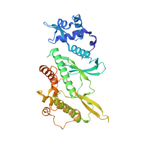
ATPase-dependent role of the atypical kinase Rio2 on the evolving pre-40S ribosomal subunit.

Ferreira-Cerca, S.Sagar, V.Schafer, T.Diop, M.Wesseling, A.M.Lu, H.Chai, E.Hurt, E.Laronde-Leblanc, N.

(2012) Nat Struct Mol Biol 19: 1316-1323

  • DOI: https://doi.org/10.1038/nsmb.2403
  • Primary Citation Related Structures: 
    4GYG, 4GYI

  • PubMed Abstract: 

    Ribosome synthesis involves dynamic association of ribosome-biogenesis factors with evolving preribosomal particles. Rio2 is an atypical protein kinase required for pre-40S subunit maturation. We report the crystal structure of eukaryotic Rio2-ATP-Mg(2+) complex. The active site contains ADP-Mg(2+) and a phosphoaspartate intermediate typically found in Na(+), K(+) and Ca(2+) ATPases but not protein kinases. Consistent with this finding, ctRio2 exhibits a robust ATPase activity in vitro. In vivo, Rio2 docks on the ribosome, with its active site occluded and its flexible loop positioned to interact with the pre-40S subunit. Moreover, Rio2 catalytic activity is required for its dissociation from the ribosome, a necessary step in pre-40S maturation. We propose that phosphoryl transfer from ATP to Asp257 in Rio2's active site and subsequent hydrolysis of the aspartylphosphate could be a trigger to power late cytoplasmic 40S subunit biogenesis.


  • Organizational Affiliation
    • Biochemistry Center, University of Heidelberg, Heidelberg, Germany.

Macromolecule Content 

  • Total Structure Weight: 45.35 kDa 
  • Atom Count: 2,835 
  • Modeled Residue Count: 339 
  • Deposited Residue Count: 397 
  • Unique protein chains: 1

Macromolecules

Find similar proteins by:|  3D Structure
Entity ID: 1
MoleculeChains  Sequence LengthOrganismDetailsImage
Rio2 kinase397Thermochaetoides thermophila DSM 1495Mutation(s): 0 
Gene Names: CTHT_0033330Rio2
EC: 2.7.11.1
UniProt
Find proteins for G0S5R3 (Chaetomium thermophilum (strain DSM 1495 / CBS 144.50 / IMI 039719))
Explore G0S5R3 
Go to UniProtKB:  G0S5R3
Entity Groups
Sequence Clusters30% Identity50% Identity70% Identity90% Identity95% Identity100% Identity
UniProt GroupG0S5R3
Sequence Annotations
Expand
Reference Sequence

Experimental Data & Validation

Experimental Data

  • Method: X-RAY DIFFRACTION
  • Resolution: 2.20 Å
  • R-Value Free:  0.226 (Depositor), 0.222 (DCC) 
  • R-Value Work:  0.168 (Depositor), 0.164 (DCC) 
  • R-Value Observed: 0.174 (Depositor) 
Space Group: H 3
Unit Cell:
Length ( Å )Angle ( ˚ )
a = 119.624α = 90
b = 119.624β = 90
c = 71.783γ = 120
Software Package:
Software NamePurpose
ADSCdata collection
PHENIXmodel building
PHENIXrefinement
HKL-2000data reduction
HKL-2000data scaling
PHENIXphasing

Structure Validation

View Full Validation Report


Ligand Structure Quality Assessment 


Entry History 

Deposition Data

Revision History  (Full details and data files)

  • Version 1.0: 2012-10-17
    Type: Initial release
  • Version 1.1: 2012-12-12
    Changes: Database references
  • Version 1.2: 2012-12-26
    Changes: Database references
  • Version 1.3: 2023-09-13
    Changes: Data collection, Database references, Derived calculations, Refinement description