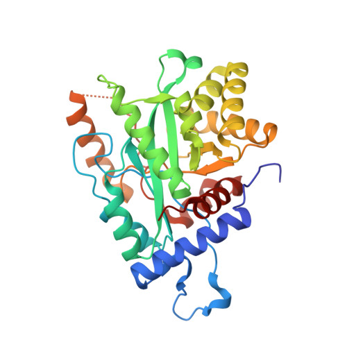
4GX8 | pdb_00004gx8

Crystal structure of a DNA polymerase III alpha-epsilon chimera


Experimental Data Snapshot

  • Method: X-RAY DIFFRACTION
  • Resolution: 1.70 Å
  • R-Value Free: 
    0.246 (Depositor), 0.261 (DCC) 
  • R-Value Work: 
    0.213 (Depositor), 0.227 (DCC) 
  • R-Value Observed: 
    0.214 (Depositor) 

Starting Model: experimental
View more details

wwPDB Validation 3D Report Full Report

Validation slider image for 4GX8

This is version 1.4 of the entry. See complete history. 

Literature

Proofreading exonuclease on a tether: the complex between the E. coli DNA polymerase III subunits alpha, {varepsilon}, theta and beta reveals a highly flexible arrangement of the proofreading domain

Ozawa, K., Horan, N.P., Robinson, A., Yagi, H., Hill, F.R., Jergic, S., Xu, Z.Q., Loscha, K.V., Li, N., Tehei, M., Oakley, A.J., Otting, G., Huber, T., Dixon, N.E.

(2013) Nucleic Acids Res 41: 5354-5367

  • DOI: https://doi.org/10.1093/nar/gkt162
  • Primary Citation Related Structures: 
    4GX8, 4GX9

  • PubMed Abstract: 

    A complex of the three (αεθ) core subunits and the β2 sliding clamp is responsible for DNA synthesis by Pol III, the Escherichia coli chromosomal DNA replicase. The 1.7 Å crystal structure of a complex between the PHP domain of α (polymerase) and the C-terminal segment of ε (proofreading exonuclease) subunits shows that ε is attached to α at a site far from the polymerase active site. Both α and ε contain clamp-binding motifs (CBMs) that interact simultaneously with β2 in the polymerization mode of DNA replication by Pol III. Strengthening of both CBMs enables isolation of stable αεθ:β2 complexes. Nuclear magnetic resonance experiments with reconstituted αεθ:β2 demonstrate retention of high mobility of a segment of 22 residues in the linker that connects the exonuclease domain of ε with its α-binding segment. In spite of this, small-angle X-ray scattering data show that the isolated complex with strengthened CBMs has a compact, but still flexible, structure. Photo-crosslinking with p-benzoyl-L-phenylalanine incorporated at different sites in the α-PHP domain confirm the conformational variability of the tether. Structural models of the αεθ:β2 replicase complex with primer-template DNA combine all available structural data.


  • Organizational Affiliation: 
    • School of Chemistry, University of Wollongong, Northfields Avenue, Wollongong, NSW 2522, Australia.

Macromolecule Content 

  • Total Structure Weight: 138.65 kDa 
  • Atom Count: 11,016 
  • Modeled Residue Count: 1,242 
  • Deposited Residue Count: 1,260 
  • Unique protein chains: 1

Macromolecules

Find similar proteins by:|  3D Structure
Entity ID: 1
MoleculeChains  Sequence LengthOrganismDetailsImage
DNA polymerase III subunit epsilon,DNA polymerase III subunit alpha
A, B, C, D
315Escherichia coli K-12Mutation(s): 1 
Gene Names: dnaQ, mutD, b0215, JW0205, dnaE, polC, b0184, JW0179
EC: 2.7.7.7
UniProt
Find proteins for P03007 (Escherichia coli (strain K12))
Explore P03007 
Go to UniProtKB:  P03007
Find proteins for P10443 (Escherichia coli (strain K12))
Explore P10443 
Go to UniProtKB:  P10443
Entity Groups
Sequence Clusters30% Identity50% Identity70% Identity90% Identity95% Identity100% Identity
UniProt GroupsP03007P10443
Sequence Annotations
Expand
Reference Sequence

Experimental Data & Validation

Experimental Data

  • Method: X-RAY DIFFRACTION
  • Resolution: 1.70 Å
  • R-Value Free:  0.246 (Depositor), 0.261 (DCC) 
  • R-Value Work:  0.213 (Depositor), 0.227 (DCC) 
  • R-Value Observed: 0.214 (Depositor) 
Space Group: P 1 21 1
Unit Cell:
Length ( Å )Angle ( ˚ )
a = 84.47α = 90
b = 56.63β = 93.52
c = 138.01γ = 90
Software Package:
Software NamePurpose
MAR345dtbdata collection
PHENIXmodel building
REFMACrefinement
MOSFLMdata reduction
SCALAdata scaling
PHENIXphasing

Structure Validation

View Full Validation Report


Entry History 

Deposition Data

Revision History  (Full details and data files)

  • Version 1.0: 2013-04-03
    Type: Initial release
  • Version 1.1: 2013-04-24
    Changes: Database references
  • Version 1.2: 2013-06-26
    Changes: Database references
  • Version 1.3: 2017-06-21
    Changes: Advisory
  • Version 1.4: 2023-11-08
    Changes: Data collection, Database references, Derived calculations, Refinement description