4GWT

Structure of racemic Pin1 WW domain cocrystallized with DL-malic acid


Experimental Data Snapshot

  • Method: X-RAY DIFFRACTION
  • Resolution: 2.25 Å
  • R-Value Free: 0.260 
  • R-Value Work: 0.238 
  • R-Value Observed: 0.239 

wwPDB Validation   3D Report Full Report


This is version 1.3 of the entry. See complete history


Literature

Evidence for small-molecule-mediated loop stabilization in the structure of the isolated Pin1 WW domain.

Mortenson, D.E.Kreitler, D.F.Yun, H.G.Gellman, S.H.Forest, K.T.

(2013) Acta Crystallogr D Biol Crystallogr 69: 2506-2512

  • DOI: https://doi.org/10.1107/S090744491302444X
  • Primary Citation of Related Structures:  
    4GWT, 4GWV

  • PubMed Abstract: 

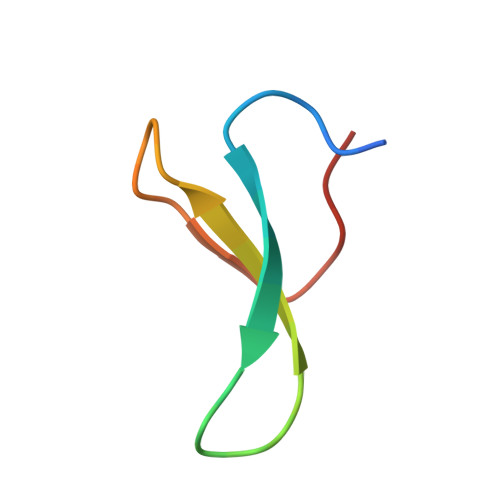
    The human Pin1 WW domain is a small autonomously folding protein that has been useful as a model system for biophysical studies of β-sheet folding. This domain has resisted previous attempts at crystallization for X-ray diffraction studies, perhaps because of intrinsic conformational flexibility that interferes with the formation of a crystal lattice. Here, the crystal structure of the human Pin1 WW domain has been obtained via racemic crystallization in the presence of small-molecule additives. Both enantiomers of a 36-residue variant of the Pin1 WW domain were synthesized chemically, and the L- and D-polypeptides were combined to afford diffracting crystals. The structural data revealed packing interactions of small carboxylic acids, either achiral citrate or a D,L mixture of malic acid, with a mobile loop region of the WW-domain fold. These interactions with solution additives may explain our success in crystallization of this protein racemate. Molecular-dynamics simulations starting from the structure of the Pin1 WW domain suggest that the crystal structure closely resembles the conformation of this domain in solution. The structural data presented here should provide a basis for further studies of this important model system.


  • Organizational Affiliation

    Department of Chemistry, University of Wisconsin-Madison, Madison, WI 53706, USA.


Macromolecules
Find similar proteins by:  (by identity cutoff)  |  3D Structure
Entity ID: 1
MoleculeChains Sequence LengthOrganismDetailsImage
Peptidyl-prolyl cis-trans isomerase NIMA-interacting 136Homo sapiensMutation(s): 0 
EC: 5.2.1.8
UniProt & NIH Common Fund Data Resources
Find proteins for Q13526 (Homo sapiens)
Explore Q13526 
Go to UniProtKB:  Q13526
PHAROS:  Q13526
GTEx:  ENSG00000127445 
Entity Groups  
Sequence Clusters30% Identity50% Identity70% Identity90% Identity95% Identity100% Identity
UniProt GroupQ13526
Sequence Annotations
Expand
  • Reference Sequence
Small Molecules
Ligands 1 Unique
IDChains Name / Formula / InChI Key2D Diagram3D Interactions
LMR
Query on LMR

Download Ideal Coordinates CCD File 
B [auth A](2S)-2-hydroxybutanedioic acid
C4 H6 O5
BJEPYKJPYRNKOW-REOHCLBHSA-N
Experimental Data & Validation

Experimental Data

  • Method: X-RAY DIFFRACTION
  • Resolution: 2.25 Å
  • R-Value Free: 0.260 
  • R-Value Work: 0.238 
  • R-Value Observed: 0.239 
  • Space Group: I 41/a
Unit Cell:
Length ( Å )Angle ( ˚ )
a = 65.985α = 90
b = 65.985β = 90
c = 38.599γ = 90
Software Package:
Software NamePurpose
DENZOdata reduction
SCALEPACKdata scaling
REFMACrefinement
PDB_EXTRACTdata extraction
PROTEUM PLUSdata collection
HKL-2000data reduction
PHASERphasing

Structure Validation

View Full Validation Report


Entry History 

Deposition Data

Revision History  (Full details and data files)

  • Version 1.0: 2013-10-16
    Type: Initial release
  • Version 1.1: 2014-01-08
    Changes: Database references
  • Version 1.2: 2017-11-15
    Changes: Refinement description
  • Version 1.3: 2023-09-13
    Changes: Data collection, Database references, Refinement description