 Download Mendeley
Northeast Structural Genomics Consortium Target OR248
Kuzin, A., Lew, S., Seetharaman, J., Rajagopalan, S., Everett, J.K., Acton, T.B., Montelione, G.T., Tong, L., Hunt, J.F.To be published.
Experimental Data Snapshot
Starting Model: experimental
View more details
wwPDB Validation 3D Report Full Report

Entity ID: 1 | |||||
|---|---|---|---|---|---|
| Molecule | Chains | Sequence Length | Organism | Details | Image |
| De novo designed serine hydrolase | 199 | N/A | Mutation(s): 0 | 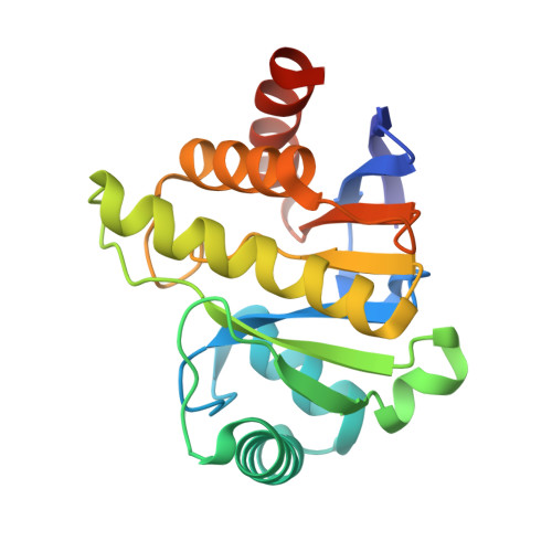 | |
Entity Groups | |||||
| Sequence Clusters | 30% Identity50% Identity70% Identity90% Identity95% Identity100% Identity | ||||
Sequence AnnotationsExpand | |||||
Reference Sequence | |||||
| Ligands 3 Unique | |||||
|---|---|---|---|---|---|
| ID | Chains | Name / Formula / InChI Key | 2D Diagram | 3D Interactions | |
| MES Download:Ideal Coordinates CCD File | C [auth A] | 2-(N-MORPHOLINO)-ETHANESULFONIC ACID C6 H13 N O4 S SXGZJKUKBWWHRA-UHFFFAOYSA-N | 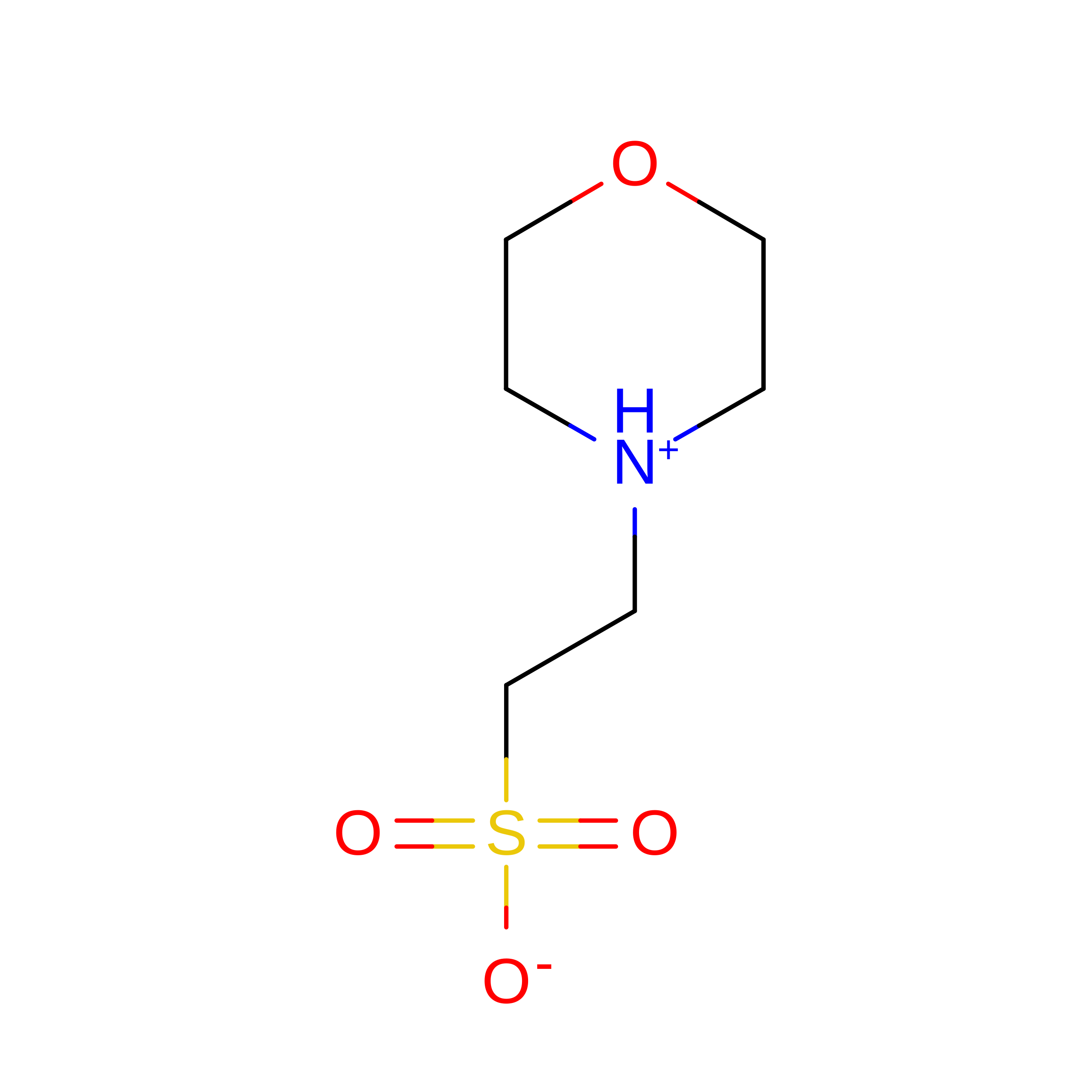 | ||
| PO4 Download:Ideal Coordinates CCD File | B [auth A] | PHOSPHATE ION O4 P NBIIXXVUZAFLBC-UHFFFAOYSA-K |  | ||
| ACY Download:Ideal Coordinates CCD File | D [auth A] | ACETIC ACID C2 H4 O2 QTBSBXVTEAMEQO-UHFFFAOYSA-N | 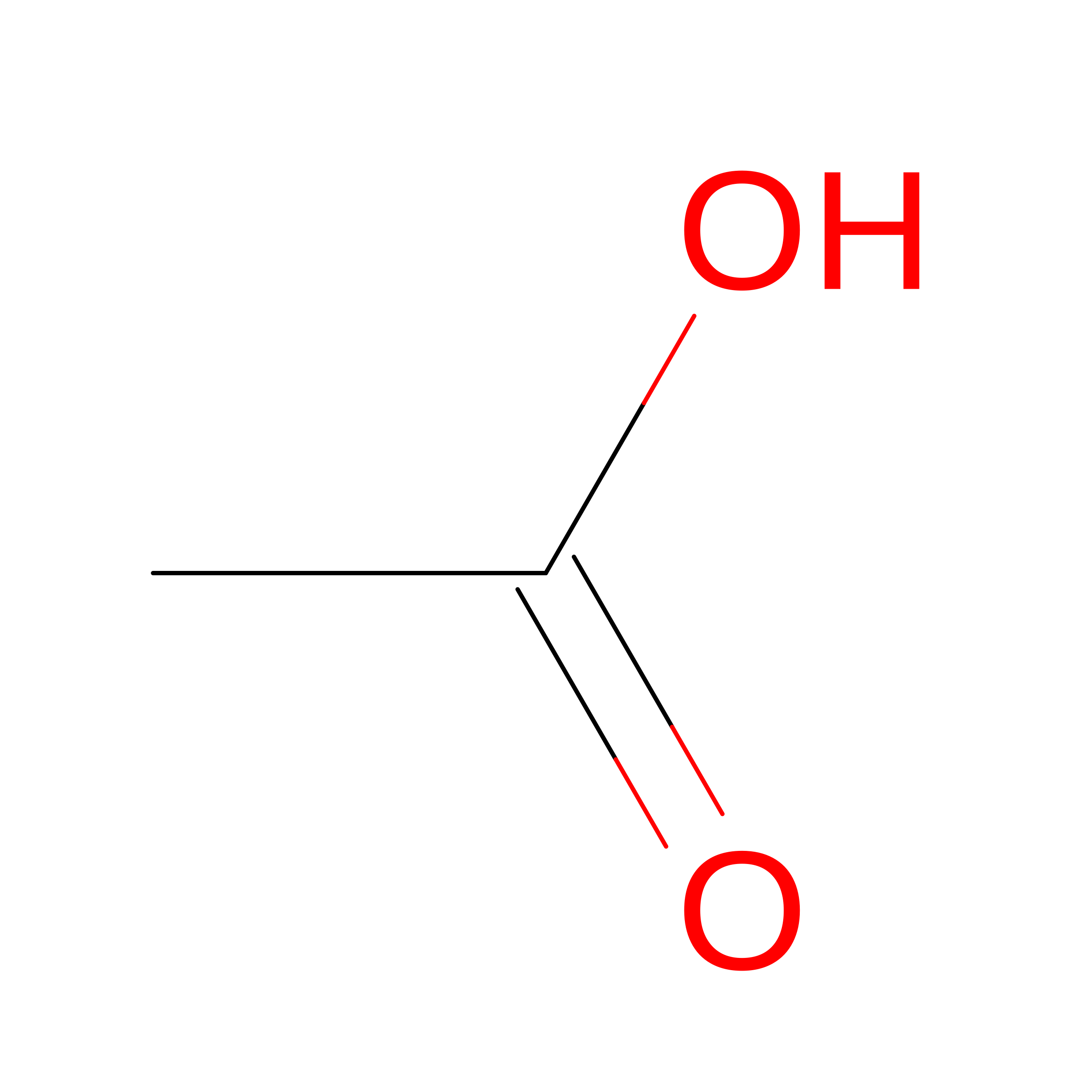 | ||
| Length ( Å ) | Angle ( ˚ ) |
|---|---|
| a = 55.644 | α = 90 |
| b = 62.052 | β = 90 |
| c = 107.067 | γ = 90 |
| Software Name | Purpose |
|---|---|
| PHENIX | refinement |
| PDB_EXTRACT | data extraction |
| msr165 | data collection |
| HKL-2000 | data reduction |
| HKL-2000 | data scaling |
| BALBES | phasing |
