


Crystal structure of AKR1B10 complexed with NADP+ and Caffeic acid phenethyl ester
Liping, Z., Xuehua, Z., Shangke, C., Jing, Z.To be published.
Experimental Data Snapshot
Starting Model: experimental
View more details
Entity ID: 1 | |||||
|---|---|---|---|---|---|
| Molecule | Chains | Sequence Length | Organism | Details | Image |
| Aldo-keto reductase family 1 member B10 | 318 | Homo sapiens | Mutation(s): 0 Gene Names: AKR1B10 EC: 1.1.1 (PDB Primary Data), 1.1.1.372 (UniProt), 1.1.1.21 (UniProt), 1.1.1.300 (UniProt), 1.1.1.54 (UniProt) | 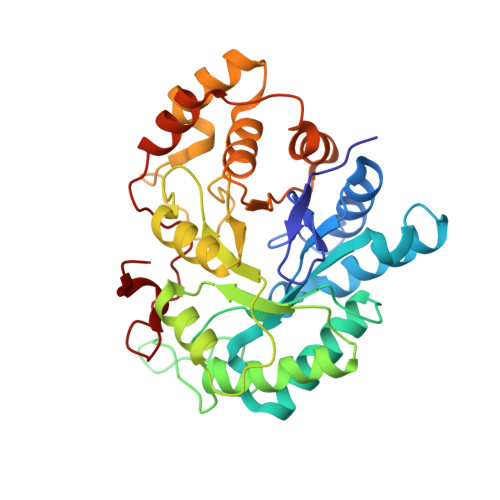 | |
UniProt & NIH Common Fund Data Resources | |||||
PHAROS: P15121 GTEx: ENSG00000085662 | |||||
Entity Groups | |||||
| Sequence Clusters | 30% Identity50% Identity70% Identity90% Identity95% Identity100% Identity | ||||
| UniProt Group | P15121 | ||||
Sequence AnnotationsExpand | |||||
Reference Sequence | |||||
| Ligands 2 Unique | |||||
|---|---|---|---|---|---|
| ID | Chains | Name / Formula / InChI Key | 2D Diagram | 3D Interactions | |
| NAP Download:Ideal Coordinates CCD File | C [auth A] | NADP NICOTINAMIDE-ADENINE-DINUCLEOTIDE PHOSPHATE C21 H28 N7 O17 P3 XJLXINKUBYWONI-NNYOXOHSSA-N |  | ||
| QAP Download:Ideal Coordinates CCD File | B [auth A] | 2-phenylethyl (2E)-3-(3,4-dihydroxyphenyl)prop-2-enoate C17 H16 O4 SWUARLUWKZWEBQ-VQHVLOKHSA-N |  | ||
| Length ( Å ) | Angle ( ˚ ) |
|---|---|
| a = 90.593 | α = 90 |
| b = 90.593 | β = 90 |
| c = 78.372 | γ = 120 |
| Software Name | Purpose |
|---|---|
| CrysalisPro | data collection |
| MOLREP | phasing |
| PHENIX | refinement |
| CrysalisPro | data reduction |
| SCALA | data scaling |


