4GP2 | pdb_00004gp2

Crystal structure of ISOPRENOID SYNTHASE A3MSH1 (TARGET EFI-501992) from pyrobaculum calidifontis complexed with DMAPP and Magnesium


Experimental Data Snapshot

  • Method: X-RAY DIFFRACTION
  • Resolution: 2.00 Å
  • R-Value Free: 
    0.207 (Depositor), 0.208 (DCC) 
  • R-Value Work: 
    0.171 (Depositor), 0.172 (DCC) 
  • R-Value Observed: 
    0.172 (Depositor) 

Starting Model: experimental
View more details

wwPDB Validation 3D Report Full Report

Validation slider image for 4GP2

Ligand Structure Quality Assessment 


This is version 1.2 of the entry. See complete history

Literature

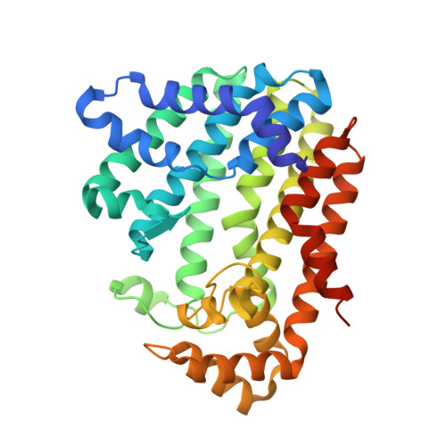
Crystal Structure of Isoprenoid Synthase from Pyrobaculum Calidifontis

Patskovsky, Y.Toro, R.Bhosle, R.Hillerich, B.Seidel, R.D.Washington, E.Scott Glenn, A.Chowdury, S.Evans, B.Hammonds, J.Zencheck, W.D.Imker, H.J.Poulter, C.D.Gerlt, J.A.Almo, S.C.

To be published.

Macromolecule Content 

  • Total Structure Weight: 81.43 kDa 
  • Atom Count: 5,607 
  • Modeled Residue Count: 673 
  • Deposited Residue Count: 716 
  • Unique protein chains: 1

Macromolecules

Find similar proteins by:|  3D Structure
Entity ID: 1
MoleculeChains  Sequence LengthOrganismDetailsImage
Polyprenyl synthetase
A, B
358Pyrobaculum calidifontis JCM 11548Mutation(s): 0 
Gene Names: Pcal_0150
UniProt
Find proteins for A3MSH1 (Pyrobaculum calidifontis (strain DSM 21063 / JCM 11548 / VA1))
Explore A3MSH1 
Go to UniProtKB:  A3MSH1
Entity Groups
Sequence Clusters30% Identity50% Identity70% Identity90% Identity95% Identity100% Identity
UniProt GroupA3MSH1
Sequence Annotations
Expand
Reference Sequence

Small Molecules

Ligands 2 Unique
IDChains Name / Formula / InChI Key2D Diagram3D Interactions
DMA

Query on DMA


Download:Ideal Coordinates CCD File
E [auth A],
F [auth A],
K [auth B],
L [auth B]
DIMETHYLALLYL DIPHOSPHATE
C5 H12 O7 P2
CBIDRCWHNCKSTO-UHFFFAOYSA-N
MG

Query on MG


Download:Ideal Coordinates CCD File
C [auth A]
D [auth A]
G [auth A]
H [auth B]
I [auth B]
C [auth A],
D [auth A],
G [auth A],
H [auth B],
I [auth B],
J [auth B]
MAGNESIUM ION
Mg
JLVVSXFLKOJNIY-UHFFFAOYSA-N

Experimental Data & Validation

Experimental Data

  • Method: X-RAY DIFFRACTION
  • Resolution: 2.00 Å
  • R-Value Free:  0.207 (Depositor), 0.208 (DCC) 
  • R-Value Work:  0.171 (Depositor), 0.172 (DCC) 
  • R-Value Observed: 0.172 (Depositor) 
Space Group: P 21 21 21
Unit Cell:
Length ( Å )Angle ( ˚ )
a = 53.991α = 90
b = 93.559β = 90
c = 146.886γ = 90
Software Package:
Software NamePurpose
PHASERphasing
REFMACrefinement
HKL-2000data reduction
HKL-2000data scaling

Structure Validation

View Full Validation Report


Ligand Structure Quality Assessment 


Entry History 

Revision History  (Full details and data files)

  • Version 1.0: 2012-10-10
    Type: Initial release
  • Version 1.1: 2018-01-24
    Changes: Structure summary
  • Version 1.2: 2023-09-13
    Changes: Data collection, Database references, Derived calculations, Refinement description